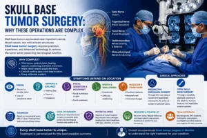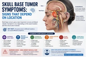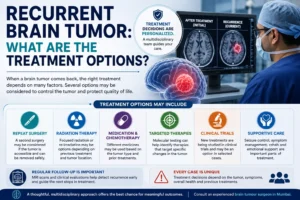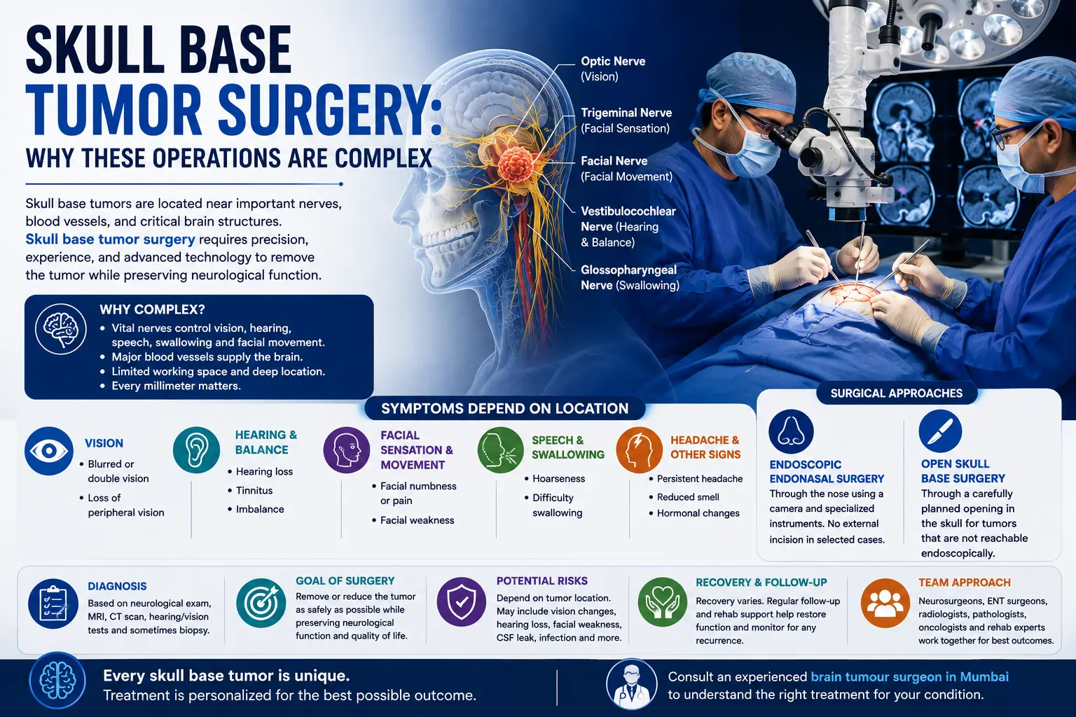
The skull base is one of the most crowded areas of the nervous system. Important arteries, veins, cranial nerves, the brainstem, the eyes, the inner ear, the nose, and the upper spinal cord can all lie close to a tumor. For this reason, skull base tumor surgery is rarely about removing a mass alone. The surgeon must also protect the structures responsible for vision, hearing, facial movement, swallowing, speech, balance, and other neurological functions.
Some skull base tumors can be reached through the nose with an endoscope. Others require a carefully planned opening in the skull, and some are better treated with radiation or observation rather than surgery. The appropriate approach depends on the exact diagnosis, tumor location, size, growth, symptoms, imaging findings, and individual health.
Why Is Skull Base Tumor Surgery Complex?
Skull base tumor surgery is complex because tumors often grow beside or around important cranial nerves and major blood vessels in an area that is difficult to access. The surgical route must provide enough access to treat the tumor while avoiding unnecessary movement or injury to the brain, nerves, arteries, and other normal structures.
The skull base forms the floor beneath the brain. It also contains multiple openings through which nerves and blood vessels travel between the brain, face, neck, eyes, ears, and throat. Johns Hopkins describes this region as a crowded and complex area that can be difficult to see and reach surgically.
A tumor may be close to structures controlling:
- Vision and eye movement
- Hearing and balance
- Facial sensation
- Facial movement
- Swallowing
- Voice
- Tongue movement
- Hormone function
- Blood supply to the brain
This means two skull base tumors of the same size can require very different operations.
What Types of Tumors May Require Skull Base Surgery?
The term “skull base tumor” covers many different diagnoses rather than one disease.
Examples include:
- Meningiomas
- Pituitary tumors
- Vestibular schwannomas
- Chordomas
- Chondrosarcomas
- Craniopharyngiomas
- Certain sinonasal tumors
- Other benign or malignant lesions
Some grow from the skull-base bones, while others arise from nerves, meninges, the pituitary region, or tissues of the nose and sinuses.
This difference matters because treatment for a pituitary tumor can be very different from treatment for a chordoma or a skull-base meningioma.
Mayo Clinic notes that treatment planning considers the tumor’s location, growth, spread, symptoms, tumor-cell testing, age, and general health.
When Is Skull Base Tumor Surgery Considered?
Surgery may be considered when removing or reducing the tumor is expected to provide a meaningful benefit.
Possible reasons include:
- Progressive neurological symptoms
- Compression of cranial nerves
- Threatened vision or hearing
- Pressure on the brainstem or brain
- Continued tumor growth on follow-up imaging
- A tumor that can be safely removed
- A need for tissue diagnosis
- A malignant tumor where surgical removal forms part of treatment
But surgery is not mandatory for every skull base lesion.
Small tumors that are stable and causing no significant symptoms may sometimes be monitored with MRI. Radiation therapy can also be an alternative or additional treatment depending on tumor type and location. Johns Hopkins describes observation, surgery, and radiation as possible components of skull-base tumor management.
How Do Surgeons Decide Which Route to Use?
There is no single operation called “skull base surgery.”
The approach is chosen according to where the tumor sits and what structures lie between the surgeon and the lesion.
Broadly, the options include:
Endoscopic Endonasal Surgery
An endoscopic endonasal approach reaches selected tumors through the nostrils and sinuses.
A thin camera called an endoscope provides a magnified view, while specialised instruments are used to work at the base of the skull.
This approach may be suitable for selected midline skull-base tumors such as certain:
- Pituitary tumors
- Chordomas
- Craniopharyngiomas
- Meningiomas
The main advantage is that the surgeon may reach the lesion without a large scalp incision or a traditional open route through the skull.
However, “minimally invasive” does not mean simple or risk-free. The operation can still take place very close to the carotid arteries, optic nerves, pituitary structures, and brain.
Open Skull Base Surgery
Some tumors are better approached through a craniotomy, where a section of skull is temporarily removed.
Different open routes may be used depending on whether the tumor is:
- Behind the eye
- Beside the brainstem
- Near the inner ear
- Around major arteries
- Extending laterally beyond the reach of a nasal approach
Examples include retrosigmoid, far-lateral, supraorbital, and orbitozygomatic approaches. The chosen route aims to provide access while limiting unnecessary retraction or disturbance of normal brain tissue.
Combined Approaches
Some particularly extensive lesions may require more than one route, either during the same treatment period or in stages.
Mayo Clinic notes that patient-specific skull-base care may involve minimally invasive, open, endoscopic, or combined procedures depending on anatomy.
Why Is Complete Cranial Base Tumor Removal Not Always the Goal?
Patients understandably want to hear that the entire tumor will be removed. In some situations that is achievable and appropriate.
In others, attempting complete cranial base tumor removal may create more harm than leaving a small amount behind.
A tumor may be:
- Wrapped around a major artery
- Adherent to the optic nerve
- Closely attached to the facial nerve
- Invading the cavernous sinus
- Pressing on the brainstem
- Surrounding several lower cranial nerves
Trying to separate every visible fragment can sometimes risk permanent neurological injury.
Mayo Clinic describes the goal of skull-base treatment as removing or controlling the tumor while avoiding damage to nearby tissues. For some tumors, partial removal followed by observation or radiation may therefore represent the safer strategy.
The best operation is not always the one that removes the most tissue. It is the one that provides appropriate tumor control while protecting neurological function as far as possible.
How Is Complex Brain Tumor Surgery Planned?
Detailed planning is particularly important before complex brain tumor surgery at the skull base.
MRI
MRI shows the tumor and its relationship with:
- Brain tissue
- Cranial nerves
- Brainstem
- Blood vessels
- Optic pathways
- Pituitary region
Contrast-enhanced MRI may help define tumor boundaries and vascular relationships.
CT
CT provides detailed information about bone.
It may be particularly helpful for tumors that arise from, destroy, thicken, or extend through skull-base bone.
Vascular Imaging
If a tumor lies close to major arteries or veins, CT angiography, MR angiography, or catheter angiography may be used in selected patients.
Hearing, Vision or Hormone Testing
Depending on location, the preoperative evaluation may include:
- Audiometry
- Visual-field testing
- Eye-movement assessment
- Endocrine blood tests
- Swallowing or voice evaluation
Advanced imaging, navigation systems, 3D models, and intraoperative imaging can also assist surgical planning in selected cases.
Why Does Skull Base Surgery Often Require a Team?
The anatomy crosses traditional medical specialties.
Depending on the tumor, the surgical or treatment team may include:
- Neurosurgery
- ENT or rhinology/skull-base surgery
- Head and neck surgery
- Neuroradiology
- Ophthalmology
- Endocrinology
- Radiation oncology
- Medical oncology
- Pathology
For an endoscopic nasal approach, for example, an ENT skull-base surgeon and neurosurgeon may work together. Other tumors may require specialists in hearing, eye function, vascular surgery, or oncology.
Both Mayo Clinic and Johns Hopkins describe multidisciplinary care as an important part of complex skull-base treatment.
What Are the Possible Risks of Skull Base Tumor Surgery?
The risks are determined largely by location.
General surgical risks may include:
- Bleeding
- Infection
- Blood clots
- Anaesthetic complications
- Seizures
- Brain swelling
Location-specific complications may include:
- Vision loss or double vision
- Hearing changes
- Facial weakness
- Facial numbness
- Balance problems
- Swallowing difficulty
- Voice changes
- Hormonal problems
- Injury to major blood vessels
Cerebrospinal Fluid Leak
Cerebrospinal fluid, or CSF, is the clear fluid surrounding the brain and spinal cord.
Because skull-base operations can create an opening between the intracranial space and the nose or sinuses, CSF leakage is a recognised concern after certain procedures. Skull-base reconstruction techniques are used when needed to close this pathway. Untreated CSF leaks can increase the risk of infection, including meningitis.
The relevant risks should be discussed according to the patient’s actual MRI and proposed approach rather than from a generic complication list.
What Is Recovery Like After Skull Base Surgery?
Recovery varies widely because the term skull-base surgery includes many different procedures.
A small endoscopic operation through the nose is very different from an extensive open operation near the brainstem.
Early hospital care may involve monitoring:
- Neurological function
- Vision
- Eye movements
- Facial movement
- Hearing
- Swallowing
- Fluid balance
- Wound or nasal healing
Some patients may need:
- Physiotherapy
- Balance rehabilitation
- Speech or swallowing therapy
- Hearing rehabilitation
- Hormone replacement
- Follow-up with ophthalmology or ENT
The recovery period depends on tumor type, surgical route, extent of surgery, preoperative neurological deficits, complications, age, and overall health.
Follow-up MRI is also important because residual or recurrent tumor may need continued monitoring or additional treatment.
Which Symptoms After Surgery Need Prompt Medical Attention?
The surgeon’s own discharge instructions should always take priority.
Urgent medical review may be appropriate for:
- New weakness or numbness
- Sudden or worsening vision problems
- New facial weakness
- Significant new difficulty swallowing
- A seizure
- Loss of consciousness
- Increasing confusion
- Severe or rapidly worsening headache
- Persistent clear watery drainage from the nose after endonasal surgery
- Fever with neurological or wound symptoms
- Significant wound swelling or discharge
New neurological problems after skull-base surgery should not be ignored or assumed to be part of normal recovery.
When Should You Consult a Neurosurgeon?
A neurosurgical consultation becomes relevant when MRI or CT identifies a skull-base tumor that may require surgery, biopsy, monitoring, or multidisciplinary treatment.
A skull base neurosurgeon may review:
- The actual MRI and CT images
- Tumor type and likely diagnosis
- Cranial nerves involved
- Major arteries and veins nearby
- Whether surgery is necessary
- Whether an endoscopic or open approach is more suitable
- How much tumor can realistically be removed
- Which neurological functions are at risk
- Whether radiation is an alternative or additional treatment
- Expected recovery and follow-up
Patients with complex skull-base lesions can seek assessment from a brain tumor surgeon in Mumbai to review the imaging and discuss the available approaches.
Dr. Mazda K. Turel can assess the relationship between the lesion, neurological symptoms, and surrounding anatomy before discussing whether surgery or another treatment strategy may be appropriate.
Conclusion
Skull base tumor surgery is complex because the surgeon is operating in a confined region containing structures essential for vision, hearing, facial movement, swallowing, brain function, and blood supply. The difficulty is not simply reaching the tumor—it is deciding how to treat it without creating unnecessary neurological harm.
MRI, CT, cranial nerve testing, and detailed surgical planning help determine whether an endoscopic, open, combined, or non-surgical approach is most suitable. Complete removal may be possible for some tumors, while partial removal followed by monitoring or radiation may be safer for others.
A useful surgical plan therefore considers more than tumor size. Diagnosis, location, growth, symptoms, nearby nerves and vessels, overall health, and the likely consequences of treatment all need to be weighed together.
Frequently Asked Questions
Why is skull base tumor surgery considered difficult?
Skull base tumor surgery is difficult because the skull base contains many cranial nerves, major arteries, veins, the brainstem, and other important structures in a compact space. Tumors may also be difficult to reach. The surgeon must select a route that provides sufficient access while minimising injury to normal neurological structures.
Can all skull base tumors be removed through the nose?
No. Endoscopic endonasal surgery is useful for selected tumors that can be reached through the nose and sinuses, particularly some midline skull-base lesions. Tumors extending laterally, behind certain blood vessels, around the ear, or into other regions may require an open approach or a combination of approaches.
Does every skull base tumor need surgery?
No. Small, stable tumors that are not causing significant symptoms may sometimes be observed. Radiation can also be appropriate for selected tumors. Treatment depends on diagnosis, size, location, growth, symptoms, age, overall health, and the risks associated with removing the lesion.
Why would a surgeon leave part of a skull base tumor behind?
A small amount may deliberately be left if it is tightly attached to an important cranial nerve, brainstem structure, or major blood vessel. Removing it aggressively could cause a neurological deficit that outweighs the benefit of complete removal. Residual tumor can sometimes be monitored or treated with radiation depending on the diagnosis.
Is endoscopic skull base surgery safer than open surgery?
Neither approach is universally safer. Endoscopic surgery can avoid a large external incision and may provide direct access to selected midline tumors, while open surgery may provide safer access to lesions located elsewhere. The safest route depends on the tumor’s anatomy, vascular relationships, size, and diagnosis.
How long does recovery take after skull base surgery?
There is no single recovery timeline. Recovery after a limited endoscopic procedure may be shorter than after a large open operation, but individual factors matter. Neurological symptoms before surgery, tumor location, cranial nerve involvement, complications, and rehabilitation needs can all affect how quickly normal activities resume.
Can radiation replace skull base surgery?
Sometimes. Radiation or stereotactic radiosurgery can be appropriate for selected tumors, residual disease, recurrent tumors, or patients in whom surgery would create excessive risk. Other tumors require tissue diagnosis or decompression that radiation cannot provide immediately. The decision depends on the exact tumor rather than the skull-base location alone.