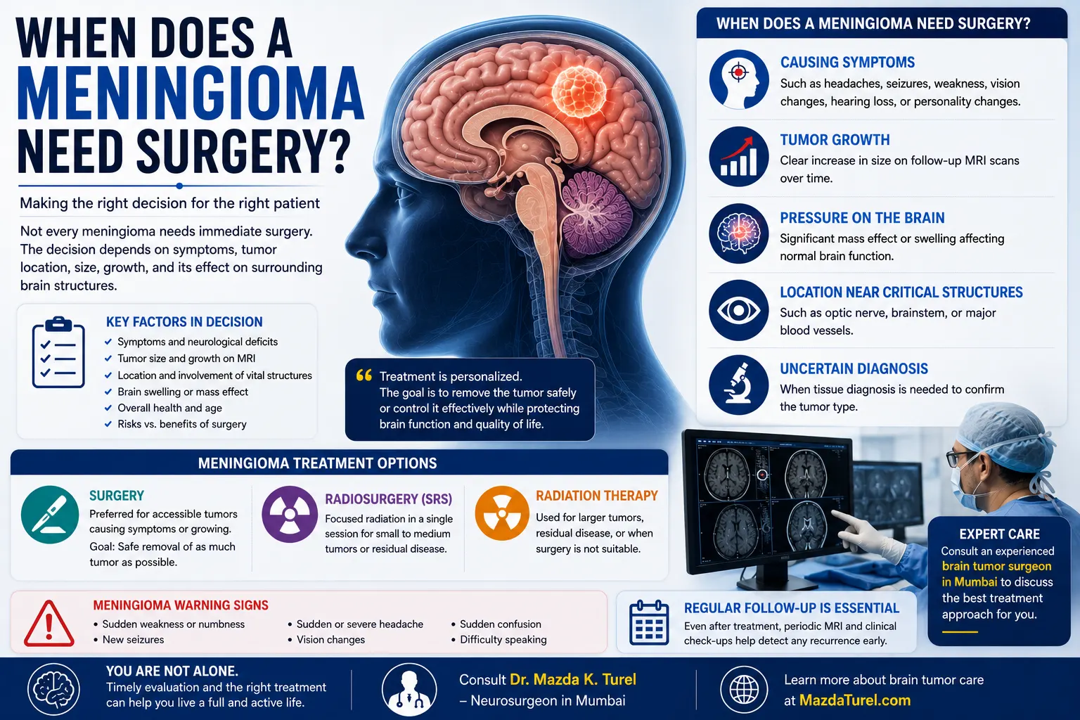
Being diagnosed with a meningioma does not automatically mean an operation is necessary. So, when does a meningioma need surgery? Surgery is generally considered when the tumor is causing neurological symptoms, shows clear growth on follow-up scans, creates pressure on the brain or nearby nerves, or is in a location where further growth could threaten functions such as vision, movement, or hearing.
Small, incidentally discovered meningiomas that are not causing symptoms may instead be monitored with regular MRI scans. Current guidance supports observation for selected asymptomatic tumors and surgery as the main treatment for meningiomas that are growing or symptomatic.
There is also no single tumor size at which every meningioma must be removed. Location, growth, symptoms, age, general health, and surgical risk all matter.
When Does a Meningioma Need Surgery Rather Than Monitoring?
A meningioma may need surgery when it causes symptoms, grows on serial MRI scans, produces significant pressure on the brain, threatens important neurological structures, or when obtaining tumor tissue is necessary for diagnosis. The decision is individual; size alone does not determine whether an operation is required.
Common meningioma surgery indications include:
- Progressive neurological symptoms
- Seizures linked to the tumor
- Weakness, numbness, or movement problems
- Progressive loss of vision or other cranial nerve problems
- Documented growth on follow-up MRI scans
- Significant surrounding brain swelling
- Pressure or displacement of nearby brain structures
- A large tumor causing clinically important mass effect
- Imaging features that make the diagnosis uncertain
- A tumor whose location makes future growth particularly concerning
The reason for surgery should always be clear. For one patient, the goal may be protecting vision. For another, it may be relieving pressure on the brain or controlling seizures. In another case, surgery may provide tissue needed to establish the exact tumor grade.
Does the Size of a Meningioma Decide Whether Surgery Is Needed?
No. There is no universal meningioma size for surgery that applies to every patient.
Size matters, but it has to be interpreted together with location and symptoms.
For example, a relatively small tumor close to the optic nerves may be clinically important if it is affecting vision. Meanwhile, a larger tumor along a less sensitive area of the brain may sometimes be found before it causes significant symptoms.
Doctors therefore assess:
- Tumor diameter and overall volume
- Location
- Growth between scans
- Brain swelling around the tumor
- Mass effect
- Relationship with cranial nerves
- Relationship with arteries and major veins
- Patient symptoms
- Age and general health
Mayo Clinic and Johns Hopkins both emphasise that treatment decisions depend on factors such as size, location, growth, symptoms, and overall health rather than size in isolation.
Growth on Follow-Up MRI Can Change the Treatment Plan
Many meningiomas are discovered unexpectedly during an MRI performed for another reason.
If the tumor is small, appears slow-growing, and is not causing symptoms, the initial approach may be active surveillance. This means repeating MRI scans and neurological reviews rather than treating the tumor immediately.
The plan can change if later scans show meaningful growth.
Doctors look at more than a single measurement. They may compare:
- Overall tumor dimensions
- Tumor volume
- New or increasing brain swelling
- Increasing pressure on surrounding structures
- New contact with important nerves or blood vessels
- Development of new symptoms
A tumor that repeatedly grows on serial imaging may be considered for treatment even before severe neurological problems develop.
Symptoms Often Matter More Than the Scan Measurement
Meningiomas can occur almost anywhere along the coverings of the brain. Their symptoms depend heavily on what lies next to them.
Symptoms that may lead a neurosurgeon to discuss treatment include:
- Seizures
- Progressive headaches associated with other neurological findings
- Weakness or numbness
- Difficulty walking
- Loss or deterioration of vision
- Double vision
- Hearing problems
- Facial numbness
- Speech difficulty
- Memory or behavioural changes
- Increasing balance problems
Meningioma symptoms can involve vision, hearing, smell, memory, speech, movement, seizures, and other functions depending on tumor location.
Symptoms should still not be attributed to a meningioma automatically. A patient can have a meningioma and a headache, for example, without the tumor necessarily being responsible for the headache.
The neurosurgeon therefore checks whether the symptoms, examination findings, and tumor location fit together.
Tumor Location Can Make Surgery More — or Less — Appropriate
Location is one of the most important factors in brain meningioma treatment.
Convexity Meningioma
Convexity meningiomas grow along the outer surface of the brain and may be relatively accessible surgically.
If one is enlarging or causing seizures, weakness, or other neurological symptoms, surgery may be considered because the tumor can sometimes be approached without passing through deep brain structures.
Skull Base Meningioma
Skull base tumors can sit close to the optic nerves, cranial nerves, brainstem, or major arteries.
In these situations, removing every visible part of the tumor may not always be the safest goal. A surgeon may deliberately leave tumor attached to a critical nerve or vessel if aggressive removal would carry an unacceptable neurological risk.
Radiation may then be considered for residual or growing tumor in selected cases.
Parasagittal Meningioma
Some meningiomas grow beside or into major venous channels responsible for draining blood from the brain.
Surgical planning must consider whether the tumor can be separated safely without compromising venous blood flow. Tumor involvement of these structures can influence how much can safely be removed.
Meningioma Near the Optic Nerves
A tumor affecting the optic nerves or nearby visual pathways deserves careful attention because progressive compression can threaten vision.
The decision may involve surgery, radiation, or a combination depending on tumor size, exact location, visual function, and treatment risk.
What Are the Alternatives to Meningioma Surgery?
The main meningioma treatment options are not limited to surgery.
Active Surveillance
Observation may be appropriate for selected meningiomas that:
- Were found incidentally
- Are not producing symptoms
- Are small or show little growth
- Are not threatening important neurological structures
Patients undergo repeat imaging and clinical review to watch for changes.
Stereotactic Radiosurgery
Stereotactic radiosurgery, or SRS, uses highly focused radiation without making a surgical incision.
It may be considered for selected smaller meningiomas, tumors in locations where conventional surgery carries greater risk, or residual tumor after an operation.
Despite its name, radiosurgery is a radiation treatment rather than an operation.
Fractionated Radiation Therapy
Radiation delivered over multiple sessions may be considered for certain tumors, particularly when their size or proximity to sensitive structures makes single-session radiosurgery less suitable.
Radiation may also be considered after surgery for residual, recurrent, or higher-grade meningiomas depending on the pathology and clinical circumstances.
What Is the Goal of Meningioma Surgery?
The goal is usually to remove as much tumor as is safely possible while preserving neurological function.
That final word matters.
Complete tumor removal may be desirable when it can be achieved without unacceptable risk. But a tumor wrapped around a major artery, cranial nerve, brainstem structure, or important venous channel may require a more conservative approach.
The operation can also provide tissue for pathological examination. This establishes the tumor diagnosis and grade more precisely than imaging alone.
The final plan may therefore involve:
- Complete removal where safely achievable
- Partial removal followed by monitoring
- Partial removal followed by radiation
- Surgery followed by radiation for selected higher-grade tumors
There is no benefit in pursuing a technically complete removal if doing so creates an unreasonable risk of permanent neurological injury.
What Are the Possible Risks of Meningioma Surgery?
Meningioma surgery is a form of brain surgery, so risks need to be discussed carefully before treatment.
General surgical risks can include:
- Bleeding
- Infection
- Seizures
- Brain swelling
- Cerebrospinal fluid leakage
- Blood clots
- Anaesthetic complications
Neurological risks depend much more on tumor location.
Surgery near visual structures may affect sight, while surgery close to areas controlling movement or speech may carry different risks. Operations involving cranial nerves can potentially affect facial movement, sensation, hearing, swallowing, or eye movement.
A neurosurgeon should explain which risks actually apply to the individual tumor rather than simply giving a general list of possible complications.
What Is Recovery Like After Meningioma Surgery?
Recovery varies considerably.
After surgery, neurological function is monitored closely, and postoperative MRI or CT imaging may be used to evaluate the surgical area and check for problems such as significant swelling or bleeding.
Some patients gradually return to normal activity over several weeks. Others need a longer recovery depending on:
- Tumor size and location
- Length and complexity of surgery
- Neurological symptoms before surgery
- Postoperative weakness or balance problems
- Seizure history
- General health
- Need for rehabilitation
Physical therapy, occupational therapy, or speech therapy may be useful if neurological functions have been affected. Recovery after meningioma surgery can range from weeks to months depending on the individual case.
When Should You Seek Urgent Medical Care?
A person with a known or suspected meningioma should seek urgent medical assessment for new severe neurological symptoms such as:
- A first seizure
- Sudden weakness or numbness
- Sudden difficulty speaking
- Sudden major vision changes
- Loss of consciousness
- Rapidly worsening confusion
- A sudden extremely severe headache
These symptoms do not necessarily mean that the meningioma has suddenly worsened. Stroke, seizure disorders, bleeding, and other neurological emergencies can produce similar signs.
When Should You Consult a Neurosurgeon?
A neurosurgical opinion is useful after a meningioma is identified, particularly if it is symptomatic, growing, large enough to create pressure, or located near critical neurological structures.
A specialist may review:
- The actual MRI images
- Tumor size and location
- Comparison with previous scans
- Brain swelling and mass effect
- Neurological symptoms
- Relationship with nerves and blood vessels
- Surgical accessibility
- Whether observation is reasonable
- Whether radiation is an alternative
- Expected surgical risks and recovery
Patients seeking specialist assessment can consult a brain tumor surgeon in Mumbai to review whether observation, surgery, radiation, or a combined approach may be appropriate.
Dr. Mazda K. Turel can review the patient’s neurological findings and imaging before discussing the balance between treating the tumor and protecting normal brain function.
Conclusion
So, when does a meningioma need surgery? The decision is rarely based on one MRI measurement. Surgery becomes more likely when the tumor is causing neurological symptoms, clearly growing, producing significant pressure, threatening vision or other important functions, or when tissue diagnosis is needed.
At the same time, many small and asymptomatic meningiomas can be monitored safely, while selected tumors may be better suited to stereotactic radiosurgery or other radiation treatment.
The safest plan comes from reviewing the actual MRI, symptoms, growth pattern, tumor location, age, general health, and risks of intervention together. The aim is not simply to remove a tumor—it is to control the disease while preserving neurological function and quality of life.
Frequently Asked Questions
What size meningioma usually needs surgery?
There is no fixed meningioma size for surgery that applies to everyone. A smaller tumor may require treatment if it is pressing on the optic nerves or another critical structure, while some larger but asymptomatic tumors may be managed differently. Size is assessed alongside symptoms, growth rate, location, brain swelling, age, and overall health.
Can a meningioma be left untreated?
Some meningiomas can be monitored rather than treated immediately. Observation is commonly considered for selected incidental, asymptomatic tumors that are not showing concerning growth or threatening critical structures. Regular MRI scans are used to detect changes. If the tumor begins growing or symptoms develop, active treatment may then be considered.
Does a growing meningioma always need surgery?
Not always. Documented growth makes treatment more likely, but surgery is only one option. Tumor location, size, symptoms, age, health, and suitability for radiation all influence the choice. Some growing tumors may be treated with stereotactic radiosurgery or fractionated radiation when surgery presents greater risk.
Is surgery necessary if a meningioma is causing seizures?
Seizures are an important symptom and should prompt specialist review, but they do not automatically mean surgery is mandatory. Doctors assess whether the tumor is likely responsible, whether seizures are controlled with medication, whether the tumor is growing, and whether it can be safely removed. These factors help determine the most appropriate treatment.
Can a skull base meningioma be completely removed?
Sometimes, but complete removal may not always be the safest option. Skull base meningiomas can surround cranial nerves, arteries, or other vital structures. A neurosurgeon may choose partial removal if aggressive surgery would threaten neurological function, with observation or focused radiation considered for the remaining tumor.
Is radiation better than surgery for a meningioma?
Neither treatment is universally better. Surgery provides immediate tumor removal and tissue diagnosis, while radiation can be useful for selected small tumors, residual disease, recurrence, or locations where surgery carries higher risk. The appropriate choice depends on tumor size, location, symptoms, pathology, age, and individual treatment risk.
Will a meningioma come back after surgery?
Recurrence is possible. The likelihood depends on tumor grade, location, how much could safely be removed, and biological characteristics. This is why follow-up MRI scans remain important even after surgery. If residual or recurrent tumor is identified, further surgery, radiation, or continued observation may be considered depending on the circumstances.