Hearing that a brain tumor is “low-grade” or “high-grade” can immediately raise questions about how quickly it may grow, whether surgery is needed, and what treatment could follow. The main difference in low grade vs high grade glioma is the biological behaviour of the tumor. Lower-grade gliomas generally grow more slowly, while higher-grade gliomas tend to grow more quickly and behave more aggressively.
Grade, however, is only part of the diagnosis. Modern glioma classification also considers the exact tumor type and molecular changes found in the tumor cells. MRI can suggest how aggressive a lesion may be, but the final classification often depends on tissue obtained through surgery or biopsy.
Understanding these differences helps patients interpret MRI reports and treatment discussions without assuming that grade alone determines the entire outlook.
Low Grade vs High Grade Glioma: What Is the Main Difference?
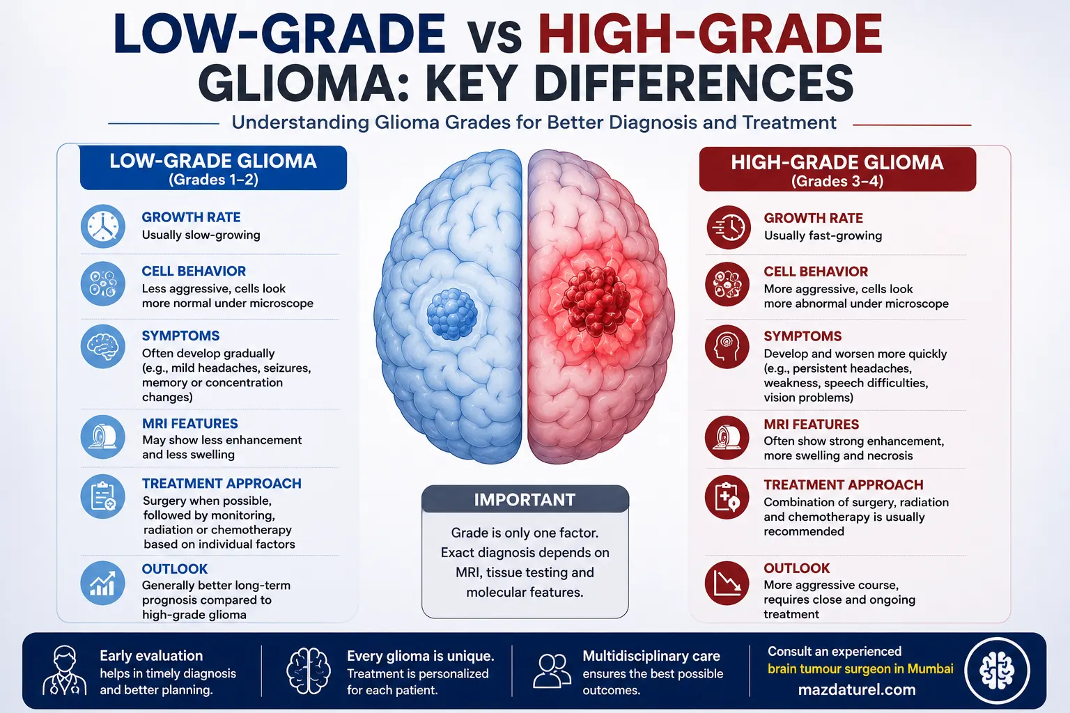
Low-grade gliomas generally grow more slowly and have less aggressive biological features, while high-grade gliomas grow faster and are more likely to invade surrounding brain tissue. Brain tumors are commonly graded from 1 to 4, but modern glioma diagnosis also uses molecular features, so grade must be interpreted together with the exact tumor type.
In broad clinical discussions:
- Grades 1 and 2 are generally considered lower grade.
- Grades 3 and 4 are generally considered higher grade.
- Grade 4 represents the most aggressive end of the grading system.
This does not mean that every grade 2 glioma behaves the same way or that every grade 3 tumor follows an identical course.
The biology of the individual tumor matters.
What Does “Grade” Mean in a Glioma?
Tumor grade describes biological features that help doctors estimate how aggressively a tumor is likely to behave.
Traditionally, pathologists look at features such as:
- How abnormal the tumor cells appear
- How actively the cells are dividing
- Evidence of tissue breakdown, called necrosis
- Abnormal blood vessel growth
- Other microscopic characteristics
Brain tumors are graded rather than staged in the way many cancers elsewhere in the body are. A grade 4 brain tumor is therefore not the same thing as “stage 4 cancer.”
For gliomas, laboratory testing now goes further than simply examining the cells under a microscope.
Why Molecular Testing Matters Alongside Glioma Grades
Modern glioma grades are interpreted together with molecular information from the tumor.
Tests may look for alterations such as:
- IDH1 or IDH2 mutations
- 1p/19q codeletion
- Other genetic or molecular changes depending on the suspected tumor
These findings help establish exactly which type of glioma is present and may provide useful information for treatment planning.
For example, current classification distinguishes different adult diffuse gliomas according to both their molecular profile and grade. Glioblastoma is a grade 4 diffuse glioma, while other gliomas can have different grades and biological characteristics.
This is why an MRI report saying “possible low-grade glioma” should not be treated as the final diagnosis.
How Does a Low-Grade Glioma Usually Behave?
A low grade glioma generally develops more slowly than a high-grade tumor.
Symptoms can therefore appear gradually, sometimes over a long period. The brain may adapt to the slowly changing environment, which can make the first signs subtle.
Possible symptoms include:
- A first seizure
- Mild or progressively changing headaches
- Difficulty concentrating
- Memory changes
- Weakness or numbness
- Problems with language
- Changes in vision
- Subtle personality or behavioural changes
A seizure is an important presentation because slowly growing tumors involving the cerebral cortex can disturb normal electrical activity.
Lower grade does not mean “harmless.”
Some low-grade diffuse gliomas infiltrate surrounding brain tissue and require treatment or long-term surveillance. Some can also change biologically over time.
How Does a High-Grade Glioma Behave Differently?
A high grade glioma generally grows more rapidly and has more aggressive tumor biology.
Symptoms may therefore progress over a shorter period.
Possible signs include:
- Increasing headaches
- New seizures
- Progressive weakness
- Speech difficulty
- Memory or behavioural changes
- Problems with balance
- Visual changes
- Nausea or vomiting
- Increasing drowsiness or confusion in more advanced cases
Glioblastoma is a specific grade 4 glioma and represents one of the most aggressive glioma types. Not every high-grade glioma is a glioblastoma.
Symptoms still cannot determine grade. A small high-grade tumor can initially cause limited symptoms, while a lower-grade tumor in an important functional area can produce significant neurological problems.
Low-Grade vs High-Grade Glioma at a Glance
Feature | Low-Grade Glioma | High-Grade Glioma |
General growth pattern | Usually slower | Usually faster |
Broad grade category | Commonly grades 1–2 | Commonly grades 3–4 |
Symptom development | May be gradual | May progress more quickly |
Brain infiltration | Depends on tumor type | Often more infiltrative/aggressive |
MRI appearance | May appear less aggressive | May show more complex aggressive features |
Tissue diagnosis | Often required | Often required |
Treatment | Depends on type, location, molecular profile and symptoms | Frequently requires combined treatment |
Follow-up | Long-term surveillance is important | Close imaging and oncological follow-up usually required |
This table gives a broad comparison only. The exact diagnosis matters more than assigning a tumor to one simple category.
Can MRI Tell Whether a Glioma Is Low Grade or High Grade?
MRI can provide valuable clues, but it cannot always determine the final grade.
A glioma MRI may show:
- Tumor location
- Tumor size and extent
- Contrast enhancement
- Surrounding brain swelling
- Pressure on nearby structures
- Areas of tissue breakdown
- Relationship with areas controlling speech, movement, vision, or other functions
High-grade tumors may show more irregular enhancement, swelling, necrosis, or other concerning features. Lower-grade gliomas may sometimes have less enhancement.
These patterns are helpful, but there is overlap.
Mayo Clinic notes that brain MRI is the imaging test used most often for glioma diagnosis, while laboratory testing of tissue helps establish the tumor type and biological characteristics.
A non-enhancing tumor is therefore not automatically low-grade, and an enhancing brain lesion is not automatically a high-grade glioma.
How Is the Final Glioma Grade Confirmed?
The most reliable classification generally comes from tumor tissue.
Tissue may be collected:
- During surgery to remove the tumor
- Through a stereotactic biopsy if immediate removal is not suitable
A neuropathologist then examines the sample and performs laboratory testing.
The results help establish:
- Whether the lesion is a glioma
- The exact glioma type
- Tumor grade
- Molecular characteristics
- Information that may influence treatment
Mayo Clinic describes tissue testing and analysis of DNA changes as important parts of confirming a glioma diagnosis and planning treatment.
Does Treatment Differ Between Low-Grade and High-Grade Glioma?
Yes, although grade is only one factor in the decision.
Treatment of Lower-Grade Glioma
Surgery is often considered when the tumor can be removed safely.
The goal is usually to remove as much tumor as possible while protecting neurological functions such as language, movement, memory, and vision.
Depending on the final diagnosis, age, remaining tumor, molecular findings and other factors, treatment after surgery may involve:
- MRI surveillance
- Radiation therapy
- Chemotherapy
- Targeted treatment in selected cases
Some lower-grade tumors may need surgery alone initially, while others require additional treatment.
Treatment of Higher-Grade Glioma
Higher-grade gliomas more commonly require a combination of treatments.
These may include:
- Maximum safe surgical removal
- Radiation therapy
- Chemotherapy
- Other drug-based or targeted treatments in selected cases
- Medicines for seizures or brain swelling
- Rehabilitation when neurological function is affected
Glioma treatment commonly begins with surgery when it can be performed safely, although radiation or systemic treatment may be needed when complete removal is not possible.
The appropriate treatment cannot be predicted from grade alone.
Why Can Glioma Surgery Be Different From Removing Other Tumors?
Many diffuse gliomas grow into surrounding brain tissue rather than forming a clean boundary that separates easily from the brain.
This creates an important surgical balance.
Removing more tumor may be beneficial, but the surgeon also needs to preserve areas responsible for:
- Movement
- Speech
- Understanding language
- Vision
- Memory
- Other essential neurological functions
For tumors close to these areas, specialised techniques such as neurological mapping or awake surgery may be considered in selected patients.
Sometimes complete removal is not safe. In such cases, removing part of the tumor or obtaining a biopsy can still provide tissue needed for diagnosis and further treatment planning.
Which Warning Signs Need Medical Attention?
Possible glioma symptoms overlap with many other neurological conditions. Medical assessment is particularly appropriate for new or progressively worsening symptoms such as:
- A first unexplained seizure
- Progressive weakness or numbness
- Increasing speech difficulty
- New visual problems
- Worsening memory or personality changes
- Headaches associated with neurological symptoms
- Increasing balance or coordination problems
Seek urgent medical care for:
- A first seizure with incomplete recovery
- Sudden weakness
- Sudden difficulty speaking
- Loss of consciousness
- Rapidly worsening confusion
- Sudden major visual changes
These symptoms can also occur with stroke, bleeding, seizures and other neurological emergencies. They should not be assumed to be caused by a glioma.
When Should You Consult a Neurosurgeon?
A neurosurgical consultation is appropriate when MRI shows a lesion suspected to be a glioma or when tissue diagnosis or tumor removal is being considered.
A neurosurgeon may review:
- The actual MRI images
- Tumor location and extent
- Whether the lesion looks infiltrative
- Neurological symptoms
- Relationship with functional brain areas
- Whether biopsy is needed
- How much tumor may be safely removed
- Risks of surgery
- How pathology may change subsequent treatment
Patients with an identified or suspected glioma may seek assessment from a brain tumor surgeon in Mumbai to understand the imaging findings and possible surgical options.
Dr. Mazda K. Turel can review the MRI alongside neurological findings before discussing whether biopsy, surgical removal, or another treatment pathway may be appropriate.
Conclusion
The central difference in low grade vs high grade glioma is how the tumor behaves biologically. Lower-grade gliomas generally grow more slowly, while higher-grade tumors tend to progress more rapidly and require more intensive treatment.
Grade alone, however, does not provide the complete diagnosis. MRI shows the tumor’s location and structural characteristics, while tissue examination and molecular testing help establish the exact glioma type and guide treatment.
For some patients, surgery may be the first step. Others may require biopsy, radiation, chemotherapy, targeted treatment or continued imaging depending on the individual tumor.
The most useful discussion therefore goes beyond asking whether a glioma is “low” or “high” grade and focuses on its precise pathology, molecular profile, location, symptoms and relationship with important areas of the brain.
Frequently Asked Questions
What is the simplest difference between low grade vs high grade glioma?
The simplest difference is growth and biological aggressiveness. A low-grade glioma generally grows more slowly, while a high-grade glioma tends to grow and infiltrate surrounding brain tissue more rapidly. However, modern diagnosis also considers tumor type and molecular features, so the final treatment plan should not be based on the grade label alone.
Is a low-grade glioma benign?
Not necessarily. Some lower-grade tumors behave relatively slowly, but diffuse low-grade gliomas can still infiltrate surrounding brain tissue and require long-term treatment or surveillance. The words “low grade” describe biological behaviour rather than guaranteeing that a tumor is harmless. The exact tumor type and molecular profile provide more useful information.
Can a low-grade glioma become high grade?
Some lower-grade gliomas can progress biologically over time, while others remain relatively slow-growing for many years. Progression is not inevitable for every tumor. The risk depends on the exact glioma type and molecular characteristics, which is why continued MRI follow-up remains important after diagnosis or treatment.
Is glioblastoma the same as high-grade glioma?
Glioblastoma is a high-grade glioma, specifically a grade 4 diffuse glioma, but it is not the only tumor included in the broader high-grade category. Not every high-grade glioma should therefore be called glioblastoma. Precise pathological and molecular classification is needed to establish the correct diagnosis.
Can MRI distinguish low-grade from high-grade glioma?
MRI can suggest whether a lesion has lower- or higher-grade characteristics, but it cannot always establish the final grade. Enhancement, necrosis, edema, growth and other imaging features provide clues. Tissue obtained through surgery or biopsy may still be required to determine the exact type, grade and molecular profile.
Do all high-grade gliomas require surgery?
Not all are safely operable. Surgery is commonly considered when useful tumor removal or tissue diagnosis can be achieved without unacceptable neurological risk. If the tumor involves critical brain areas or cannot be removed safely, biopsy, radiation, chemotherapy or other treatments may form part of the plan.
Does low grade mean treatment can be delayed?
Not automatically. Some low-grade gliomas require treatment because of their location, growth, seizures or other neurological effects. Others may be approached differently depending on the diagnosis and individual circumstances. The decision should consider MRI findings, tissue and molecular results, symptoms, age, health and the safety of surgery.