One of the first questions after being told that a glioma may need surgery is, “Can the entire tumor be removed?” The answer is not based on tumor size alone. During glioma surgery, the aim is usually to remove as much tumor as can be done safely without causing an unacceptable loss of functions such as speech, movement, vision, memory, or sensation.
This balance is often described as maximum safe resection. Gliomas can grow into surrounding brain tissue rather than forming a clean border, which means removing every abnormal-looking cell may not be possible or safe. Current neurosurgical planning therefore considers MRI findings, tumor location, neurological function, tumor biology, and the relationship of the lesion to important brain networks.
Glioma Surgery Aims for Maximum Safe Resection
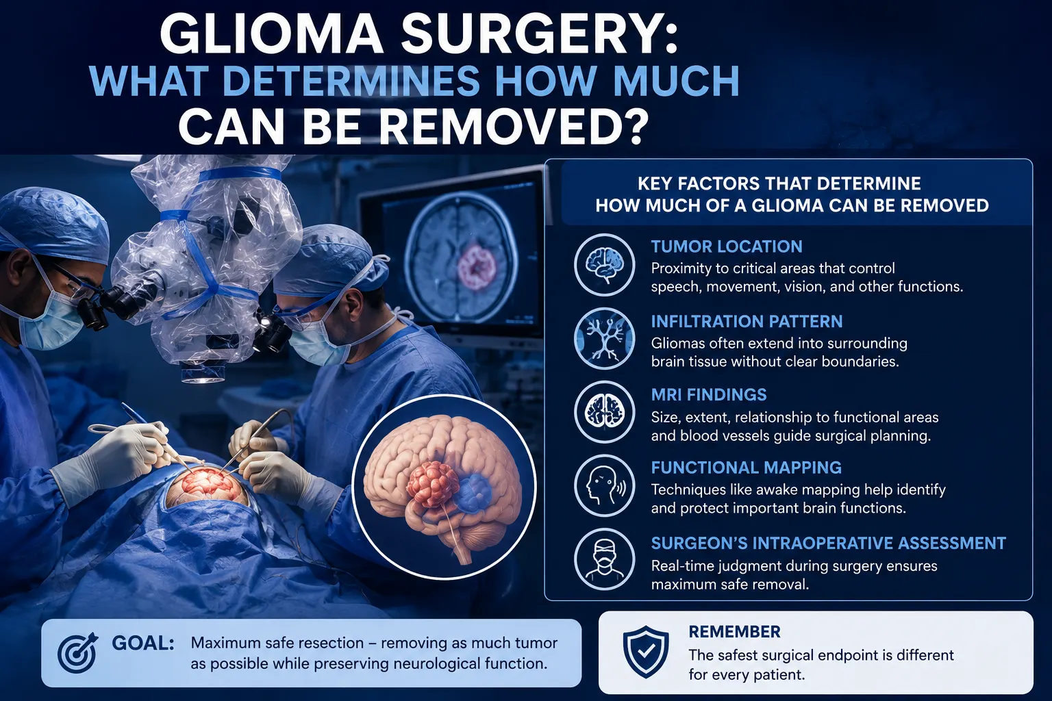
The goal of glioma surgery is generally to remove as much tumor as safely possible while preserving neurological function. How much can be removed depends on where the tumor is located, whether it infiltrates surrounding brain, how close it is to critical functional areas, and what the surgical team observes during the operation.
This is different from simply aiming for “100% removal.”
A tumor situated in a relatively accessible area may allow a larger resection. Another glioma may extend into brain tissue responsible for language, hand movement, vision, or other important functions, making aggressive removal unsafe.
The safest surgical endpoint is therefore different for every patient.
What Does “Extent of Resection” Mean?
The extent of resection in glioma describes how much visible or MRI-defined tumor has been removed during an operation.
Depending on the situation, surgeons may describe the operation as:
- Gross total resection: all tumor that can be identified using the chosen imaging criteria appears to have been removed.
- Subtotal resection: a significant amount is removed, but some tumor remains.
- Partial resection or debulking: part of the tumor is removed to reduce its volume or pressure.
- Biopsy: only a small sample is obtained mainly to establish the diagnosis.
These terms should be interpreted carefully. A “gross total resection” on postoperative MRI does not necessarily mean that every microscopic glioma cell has been eliminated.
Many diffuse gliomas infiltrate normal-looking brain beyond the visible tumor margin. NCI guidance notes that complete elimination of malignant tumors growing within brain tissue is rarely possible with surgery alone.
Tumor Location Is One of the Biggest Factors
A glioma’s location can place a practical limit on how far surgery can safely proceed.
Some areas of the brain control functions that cannot easily be replaced if damaged. These are sometimes called eloquent areas.
They may be involved in:
- Speech production
- Understanding language
- Movement
- Sensation
- Vision
- Memory
- Coordination
A tumor close to these regions may require a more cautious operation than a similar-sized tumor farther away.
For example, a glioma near the brain’s language networks may look surgically accessible on a standard MRI, but removing tissue without knowing exactly where language function lies could risk a permanent speech problem.
This is why surgical planning is based on functional anatomy as well as tumor anatomy.
Why Can Gliomas Be Difficult to Separate From Normal Brain?
Some brain tumors grow as relatively distinct masses. Diffuse gliomas can behave differently.
Tumor cells may spread between and around normal brain cells, creating a transition zone rather than a clear capsule that can simply be separated and lifted out.
This infiltrative behaviour can affect glioma removal surgery because the surgeon must decide where removing additional tissue is likely to cause more harm than benefit.
The surgical goal may therefore be to remove the visible or functionally safe portion of the tumor while leaving tissue that extends into an area essential for neurological function.
Additional treatment such as radiation therapy or chemotherapy may then be used depending on the exact diagnosis and molecular findings. NCI lists surgery, radiation therapy, chemotherapy, targeted therapy, and selected surveillance strategies among treatment approaches for adult CNS tumors.
How Does MRI Help Plan Glioma Removal?
MRI is central to planning brain tumor resection.
The neurosurgical team may review:
- Tumor size
- Exact anatomical location
- Contrast enhancement
- Surrounding swelling
- Relationship to the brain’s surface
- Involvement of deeper structures
- Nearby blood vessels
- Areas of abnormal signal extending beyond the main tumor
- Relationship with functional brain regions
The MRI also helps identify a safe route to reach the tumor.
Computer-assisted navigation can use preoperative brain images during surgery to help the surgeon understand the position of instruments relative to the tumor and nearby structures. Mayo Clinic describes computer-assisted surgery, intraoperative MRI, and awake brain surgery among techniques used to increase safe glioma removal while limiting functional injury.
What Is Awake Brain Mapping and Why Might It Be Used?
If a glioma lies close to areas controlling language or other functions, an awake craniotomy with brain mapping may be considered in selected patients.
The patient is kept comfortable with anaesthesia and sedation during appropriate parts of the operation but is awake for neurological testing when needed.
During mapping, the surgical team may ask the patient to:
- Name pictures
- Count
- Read
- Move a hand or leg
- Perform other simple tasks
The surgeon can stimulate small areas of the brain and observe whether a function is temporarily affected. This helps identify tissue that should be protected while allowing tumor removal to continue in nearby areas where it is safe.
NCI describes awake surgery with real-time neurological assessment as one method of balancing greater tumor removal against preservation of brain function.
Not every patient or glioma requires awake surgery.
Can Intraoperative Technology Help Remove More Tumor Safely?
In selected operations, additional technology may assist the surgical team.
Examples include:
Neuronavigation
Neuronavigation works somewhat like a GPS system for brain surgery. It links the patient’s anatomy to preoperative imaging so the surgeon can track the surgical position in three dimensions.
Intraoperative MRI
MRI performed during the operation can help identify tumor that may remain after the first stage of resection.
This matters because the brain can shift slightly during surgery, making the original preoperative image less accurate as the operation progresses.
Functional Mapping
Direct stimulation and other mapping methods can help define brain areas or pathways responsible for important functions.
These tools do not guarantee complete tumor removal. Their purpose is to provide more information that may help the surgeon make safer decisions during the operation. Mayo Clinic lists intraoperative MRI, awake surgery, and computer-assisted techniques among approaches used in glioma care.
Does the Glioma Type or Grade Affect the Surgical Plan?
Yes.
Surgery is planned before the final tissue diagnosis is available, but MRI appearance, age, symptoms, and other information may give the team an idea of what type of tumor is being treated.
After surgery, tissue examination provides more precise information about:
- Glioma type
- Grade
- Cellular features
- Molecular changes
These results help determine what treatment is needed next.
For example, some gliomas may be managed with surgery followed by surveillance, while others require radiation therapy, chemotherapy, or combined treatment.
For glioblastoma, NCI describes surgery followed by radiation therapy and chemotherapy as a standard treatment approach when appropriate.
The operation is therefore one part of the treatment pathway rather than the entire treatment for many diffuse gliomas.
Why Would a Surgeon Deliberately Leave Some Glioma Behind?
Leaving visible tumor can sound concerning, but there are situations where it represents the safer surgical decision.
A surgeon may stop resection if further removal would risk:
- Permanent weakness
- Loss of speech
- Difficulty understanding language
- Significant visual loss
- Major sensory problems
- Damage to important deep brain pathways
- Injury to critical blood vessels
NCI describes preservation of neurological function as an important constraint on how much CNS tumor can be surgically removed.
In these situations, treatment may continue with radiation, chemotherapy, further observation, or another strategy based on the pathology.
The objective is not simply the largest possible resection. It is the largest safe resection for that individual brain and tumor.
What Are the Possible Risks of Glioma Surgery?
The risks vary considerably according to tumor location and the type of operation.
General risks of brain surgery may include:
- Bleeding
- Infection
- Seizures
- Brain swelling
- Cerebrospinal fluid leakage
- Anaesthetic complications
Neurological risks may include:
- Weakness
- Speech or language problems
- Changes in sensation
- Vision problems
- Balance or coordination difficulties
- Cognitive changes
A tumor near the motor region creates a different risk profile from one near the visual or language networks.
This is why a useful surgical consultation should discuss the risks related to the specific MRI, rather than presenting only a generic list.
What Is Recovery Like After Glioma Surgery?
Recovery depends on the extent and location of surgery, neurological condition before the operation, age, general health, and whether any postoperative problems occur.
During the early recovery period, the medical team may monitor:
- Strength
- Speech
- Alertness
- Vision
- Seizures
- Headache
- Surgical wound
- Mobility
Postoperative MRI is commonly used to assess how much tumor remains and help guide the next phase of treatment. NCI notes that imaging may be repeated after surgery to evaluate residual tumor and plan further therapy.
Some patients recover independently, while others may need:
- Physiotherapy
- Occupational therapy
- Speech and language therapy
- Cognitive rehabilitation
Return to driving, work, exercise, and other activities should be discussed individually with the treating team.
When Should You Seek Urgent Medical Care After Surgery?
After discharge, follow the neurosurgical team’s specific instructions.
Prompt medical assessment may be necessary for:
- A new seizure
- New or worsening weakness
- New difficulty speaking
- Significant new vision changes
- Loss of consciousness
- Increasing confusion or unusual drowsiness
- Repeated vomiting
- A severe or rapidly worsening headache
- Significant wound swelling, discharge, or other signs of infection
These symptoms do not always mean a serious complication has occurred, but new neurological changes after brain surgery should not be ignored.
When Should You Consult a Neurosurgeon?
A neurosurgical consultation is particularly useful when MRI identifies a suspected glioma and there is a decision to be made between biopsy, tumor resection, or another treatment strategy.
The discussion may cover:
- What the MRI appears to show
- Whether surgery is recommended
- The realistic goal of surgery
- How much tumor may be removable
- Which brain functions are close to the lesion
- Whether awake mapping may be helpful
- Whether biopsy alone may be safer
- Possible neurological risks
- What treatment may follow surgery
Patients with a suspected or diagnosed glioma may seek evaluation from a brain tumor surgeon in Mumbai to review the actual MRI and understand the surgical options.
Dr. Mazda K. Turel can review symptoms, examination findings, and imaging before discussing the balance between tumor removal and preservation of neurological function.
Frequently Asked Questions
The central question in glioma surgery is not simply, “How much tumor can be removed?” It is, “How much can be removed safely while protecting the functions that matter to the patient’s daily life?”
Tumor location, infiltration into surrounding brain, MRI findings, functional brain areas, neurological condition, and intraoperative findings all influence the extent of resection in glioma. Techniques such as neuronavigation, functional mapping, awake surgery, and intraoperative imaging may help surgeons make more informed decisions in selected cases.
Sometimes a large resection is possible. In other cases, deliberately leaving tumor behind is the safer choice. Pathology and molecular testing then help guide radiation, chemotherapy, surveillance, or other treatment after surgery.
Frequently Asked Questions
Can a glioma be completely removed with surgery?
Sometimes the MRI-visible tumor can be completely removed, but many diffuse gliomas extend microscopically into surrounding brain tissue. A scan showing gross total resection therefore does not necessarily mean that every tumor cell has been removed. The achievable extent depends largely on tumor location, infiltration, and whether further removal would threaten neurological function.
What does maximum safe resection mean in glioma surgery?
Maximum safe resection means removing the greatest amount of tumor that can reasonably be taken out without creating unacceptable neurological harm. Surgeons balance the potential benefit of additional tumor removal against risks to speech, movement, vision, sensation, memory, and other functions. The safest endpoint is different for each patient.
Does a bigger glioma mean less of it can be removed?
Not necessarily. Size matters, but location and infiltration can be more important. A large tumor in a surgically accessible area may allow substantial removal, while a smaller glioma located within critical language or motor networks may require a more conservative approach. MRI and functional assessment help determine what is realistically removable.
What happens if only part of a glioma can be removed?
The removed tissue is analysed to establish the diagnosis and molecular characteristics. Depending on the glioma type, grade, residual tumor, age, and other factors, treatment may then include radiation therapy, chemotherapy, further monitoring, or another approach. Partial removal can still reduce tumor burden and provide tissue needed for treatment planning.
Is awake surgery necessary for every glioma?
No. Awake brain mapping is mainly considered when a tumor lies close to functional areas where direct testing during surgery could help protect neurological abilities. Other gliomas can be removed under general anaesthesia without awake testing. The decision depends on tumor location, the functions at risk, and patient suitability.
Can surgery make neurological symptoms worse?
Yes, neurological worsening is a recognised risk of brain surgery, particularly when a glioma is close to areas controlling speech, movement, vision, or other important functions. Some postoperative changes may improve as swelling settles or with rehabilitation, while others can be persistent. Individual risks should be discussed using the patient’s actual MRI.
Does glioma surgery cure the tumor?
Surgery can be an important part of treatment, but it should not automatically be described as a cure. Diffuse gliomas may extend beyond the visible surgical boundary, and many patients require additional therapy or ongoing imaging. Treatment depends on the final tissue diagnosis, molecular features, residual tumor, symptoms, and individual clinical circumstances.