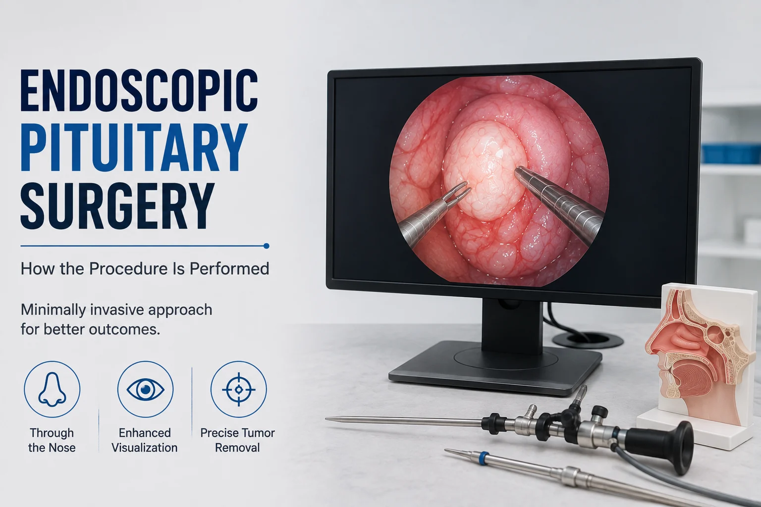
The idea of brain surgery often makes patients imagine a large scalp incision or opening in the skull. Endoscopic pituitary surgery is different. For many pituitary tumors, the surgeon reaches the gland through the natural nasal passages, passes through the sphenoid sinus behind the nose, and removes the tumor using an endoscope and specialised instruments.
This approach is also called endoscopic transnasal transsphenoidal surgery. It usually leaves no visible external scar and allows the surgeon to reach the pituitary gland from underneath the brain rather than passing through normal brain tissue.
However, surgery is not appropriate for every pituitary tumor. Treatment depends on the tumor type, hormone activity, size, relationship with the optic nerves and blood vessels, symptoms, and the patient’s overall health.
What Is Endoscopic Pituitary Surgery?
Endoscopic pituitary surgery is a minimally invasive operation in which a neurosurgeon reaches a pituitary tumor through the nose and sphenoid sinus using a thin camera called an endoscope. Special instruments are then used to remove as much tumor as can be safely reached while protecting the normal pituitary gland, optic nerves, blood vessels, and surrounding structures.
The pituitary gland sits in a small bony space called the sella turcica, located behind the nose at the base of the skull.
Because of this position, surgeons can often approach the gland directly through the nasal corridor.
The terms patients may hear include:
- Endoscopic pituitary surgery
- Endoscopic pituitary tumor removal
- Endoscopic endonasal surgery
- Transnasal surgery
- Transsphenoidal surgery
- Endoscopic transsphenoidal surgery
These terms describe closely related approaches using the nose and sphenoid sinus to access the pituitary region.
When Is Endoscopic Pituitary Surgery Considered?
Surgery is generally considered when treating the tumor is expected to provide a meaningful benefit.
Possible reasons include:
- A tumor pressing on the optic nerves or causing visual-field loss
- A nonfunctioning pituitary tumor producing pressure symptoms
- Growth on follow-up MRI
- Certain hormone-producing pituitary tumors
- Pituitary apoplexy in selected patients
- A tumor that cannot be adequately controlled with medicine
- The need to relieve pressure on nearby neurological structures
NCI guidance lists transsphenoidal surgery as an important treatment for several pituitary tumor types, including nonfunctioning, ACTH-producing, growth hormone-producing, and TSH-producing tumors. Treatment still needs to be individualised according to tumor biology and anatomy.
Prolactinomas Are an Important Exception
A prolactinoma is a pituitary tumor that produces excess prolactin.
Unlike many other pituitary tumors, prolactinomas are commonly treated first with medication. Surgery may be considered if medicine does not adequately control the condition, causes unacceptable side effects, or is unsuitable for the patient.
This is why identifying the tumor type before discussing surgery matters.
How Is Surgery Planned Before the Operation?
Good pituitary surgery begins before the patient reaches the operating theatre.
Doctors may review:
- A dedicated pituitary MRI
- Hormone blood tests
- Visual-field testing
- General blood tests
- Current medicines
- Previous pituitary treatment
- Other medical conditions
MRI helps show the tumor’s size, direction of growth, relationship with the optic chiasm, and whether it extends toward structures such as the cavernous sinus.
Hormone testing is equally important because the pituitary gland regulates several endocrine systems. Patients may be assessed by an endocrinologist before and after surgery, and visual testing may be needed when the tumor lies close to the optic pathways.
Blood-thinning medicines and other drugs may need special instructions before surgery. Patients should not stop prescribed medication unless their treating team advises them to do so.
How Is Endoscopic Pituitary Surgery Performed?
The operation is generally performed under general anaesthesia. In many centres, a neurosurgeon works together with an ear, nose and throat surgeon for the nasal and skull-base portion of the procedure.
Step 1: Entering Through the Nose
A narrow endoscope containing a camera and light is introduced through one or both nostrils.
The endoscope sends a magnified view to a monitor, allowing the surgical team to see the nasal passages and skull-base anatomy clearly.
There is usually no incision on the face or scalp.
Step 2: Reaching the Sphenoid Sinus
The surgeon guides the endoscope toward the sphenoid sinus, an air-filled space located behind the nose.
The sinus is opened to expose the bone covering the pituitary region.
Step 3: Opening the Pituitary Region
A small amount of bone over the sella turcica is removed.
The surgeon then opens the dura, the strong protective membrane covering the area, to reach the pituitary tumor.
Step 4: Removing the Tumor
Specialised instruments are passed through the nasal corridor.
The tumor is commonly removed in smaller pieces rather than being pulled out as one large mass. The surgeon works carefully around the normal pituitary tissue, optic pathways, and nearby blood vessels.
The goal is not automatically to remove every visible cell regardless of risk.
If part of a tumor is tightly attached to an important artery, nerve, or other critical structure, leaving some tissue may sometimes be safer than attempting aggressive removal.
Step 5: Reconstructing the Skull Base
After the tumor has been removed, the surgeon checks the area for cerebrospinal fluid leakage.
If necessary, tissue, fat, surgical materials, or a vascularised nasal tissue flap may be used to reconstruct the opening and reduce the risk of cerebrospinal fluid leakage.
The endoscope and instruments are then removed.
Is Pituitary Surgery Through the Nose the Same as Transsphenoidal Surgery?
Usually, yes.
Pituitary surgery through the nose describes the route, while transsphenoidal surgery means that the surgeon reaches the pituitary gland through the sphenoid sinus.
The operation can be performed with either:
- An endoscope
- A surgical microscope
Modern endoscopic pituitary tumor removal uses a camera at the tip of the endoscope to provide a close, wide view of the operative area. Mayo Clinic notes that endoscopic transsphenoidal surgery is now the most commonly performed form of the transsphenoidal approach.
What Are the Possible Risks?
Endoscopic pituitary surgery avoids a traditional craniotomy in many patients, but it is still a skull-base neurosurgical procedure and carries recognised risks.
Cerebrospinal Fluid Leak
Cerebrospinal fluid, or CSF, surrounds the brain and spinal cord.
Because the operation enters an area next to this fluid space, CSF may occasionally leak through the surgical opening and appear as clear watery drainage from the nose.
Persistent leakage may require additional treatment or another procedure.
Hormone Problems
The normal pituitary gland lies next to the tumor.
Surgery can sometimes reduce normal pituitary hormone production, known as hypopituitarism. Some patients may need temporary or long-term hormone replacement depending on which hormones are affected.
Excessive Thirst and Urination
Temporary or, less commonly, persistent arginine vasopressin deficiency—previously called central diabetes insipidus—can occur after pituitary surgery.
Typical signs include unusually large amounts of urine and intense thirst.
Sodium or Fluid Imbalance
Changes in pituitary function after surgery can disturb the body’s regulation of water and sodium.
This is one reason fluid intake, urine output, sodium levels, and hormone levels may be monitored after the operation.
Other Possible Risks
Other complications can include:
- Bleeding
- Infection or meningitis
- Nasal or sinus problems
- Reduced sense of smell
- Visual problems
- Injury to nearby blood vessels
- Anaesthetic complications
The individual risk depends on the tumor’s size, extension, previous treatment, surrounding anatomy, and the patient’s overall health.
What Happens Immediately After Surgery?
After the procedure, patients are monitored closely while recovering from anaesthesia.
The healthcare team may track:
- Blood pressure and other vital signs
- Vision
- Urine output
- Fluid intake
- Sodium levels
- Cortisol and other hormone levels
- Nasal drainage
- Headache or nausea
Hospital stay is often relatively short after uncomplicated transsphenoidal surgery. Mayo Clinic describes a typical stay of one to three days, while Johns Hopkins notes that some patients stay around one or two days. Individual recovery may be longer when more monitoring or treatment is required.
Nasal congestion, mild bloody drainage, tiredness, and headache can occur during early recovery.
Pituitary Surgery Recovery at Home
Pituitary surgery recovery involves protecting the surgical area while the tissues behind the nose and at the skull base heal.
Patients may temporarily be instructed to avoid:
- Heavy lifting
- Vigorous exercise
- Straining
- Forceful nose blowing
- Picking the nose
- Frequent bending forward
The exact restrictions and duration vary, so the surgical team’s discharge instructions should take priority.
Follow-up may include:
- Neurosurgical review
- Endocrinology assessment
- Hormone blood tests
- Repeat pituitary MRI
- Visual-field testing if vision was affected
Recovery also depends on the underlying tumor. Hormonal symptoms may require continued monitoring even if imaging shows good tumor removal.
Which Symptoms After Surgery Need Prompt Medical Attention?
Contact the treating team promptly or seek urgent medical care for symptoms such as:
- Clear, persistent watery fluid draining from the nose
- A severe or worsening headache
- Fever
- Repeated vomiting
- New or worsening vision problems
- Significant nose bleeding
- Increasing confusion or unusual drowsiness
- Markedly increased urination and thirst
- New weakness or other neurological changes
Johns Hopkins specifically advises reporting persistent headache, nausea or vomiting, fever, nasal bleeding, watery nasal discharge, and increased urination after endoscopic pituitary surgery.
When Should You Consult a Neurosurgeon?
A neurosurgical consultation is relevant when MRI shows a pituitary tumor that may require removal or decompression.
A neurosurgeon may review:
- The actual MRI images
- Tumor size and extension
- Relationship with the optic chiasm
- Cavernous sinus involvement
- Visual symptoms
- Hormone results
- Whether medication should be tried first
- Whether an endoscopic approach is suitable
- Whether complete removal can be attempted safely
- Expected risks and postoperative monitoring
Patients seeking evaluation can consult a brain tumor surgeon in Mumbai to understand whether an endoscopic or another neurosurgical approach is appropriate.
Dr. Mazda K. Turel can review imaging, symptoms, neurological findings, vision assessments, and endocrine results before discussing treatment. Pituitary tumor management often works best as a multidisciplinary process involving neurosurgery, endocrinology, radiology, and ophthalmology when required.
Conclusion
Endoscopic pituitary surgery allows many pituitary tumors to be approached through the nose and sphenoid sinus without a visible external incision or traditional craniotomy. An endoscope gives the surgical team a detailed view while specialised instruments are used to remove the tumor and relieve pressure on structures such as the optic nerves.
The procedure still requires careful planning because the pituitary gland sits close to important blood vessels, visual pathways, and hormone-producing tissue. Risks can include CSF leakage, hormonal changes, fluid imbalance, infection, bleeding, and visual problems.
The decision to operate should therefore be based on the tumor type, symptoms, hormone findings, MRI appearance, visual function, and individual treatment risks. Postoperative endocrine testing, MRI follow-up, and attention to recovery instructions remain essential parts of care.
Frequently Asked Questions
Is endoscopic pituitary surgery major brain surgery?
Endoscopic pituitary surgery is a neurosurgical procedure, but it generally reaches the pituitary through the nose rather than through a traditional opening in the skull. The approach does not require the surgeon to pass through normal brain tissue. It still carries important risks because the pituitary gland, optic nerves, carotid arteries, and skull-base structures are close together.
Will there be a visible scar after pituitary surgery through the nose?
Usually not. Endoscopic transnasal surgery is performed through the nostrils, so there is generally no visible facial or scalp incision. Some internal nasal tissue may be used during reconstruction if needed. Patients can still experience nasal stuffiness, drainage, or temporary sinus-related symptoms while the area heals.
Can every pituitary tumor be removed endoscopically?
No. Many pituitary tumors can be treated through the endonasal transsphenoidal route, but anatomy matters. Very large tumors or lesions extending around critical blood vessels, nerves, or into certain parts of the brain may not be fully removable through this approach. In selected cases, another surgical strategy or combined treatment may be more appropriate.
How long is recovery after endoscopic pituitary surgery?
Early hospital recovery is often measured in days, while return to normal activities takes longer. The exact timeline depends on tumor size, hormonal changes, complications, nasal healing, the patient’s general health, and the type of work or exercise involved. Restrictions on lifting, straining, vigorous exercise, and nose blowing commonly continue during the early healing period.
Will vision improve after pituitary tumor removal?
Vision may improve if the tumor was compressing the optic nerves or optic chiasm and that pressure is successfully relieved. However, recovery cannot be guaranteed. The outcome depends on factors such as the degree and duration of visual damage, tumor anatomy, and individual nerve recovery. Formal visual-field testing may be repeated after treatment.
Will I need hormone medicines after pituitary surgery?
Some patients do, while others do not. The normal pituitary gland may continue working adequately, or one or more hormones may temporarily or permanently become deficient. Blood tests after surgery help determine whether replacement treatment is needed. Hormone monitoring with an endocrinologist is therefore a key part of postoperative care.
Can the pituitary tumor grow back after surgery?
Residual or recurrent tumor is possible, depending on the tumor type, size, biological behaviour, and how much could safely be removed. Follow-up MRI scans and hormone tests help monitor the pituitary region over time. If tumor remains or later grows, treatment options may include further surgery, medication, radiation, or observation depending on the diagnosis.