Back or neck pain often leads to an MRI, and the report may contain terms such as “disc bulge,” “disc protrusion,” or “disc herniation.” These words can sound similar, but they do not always describe the same change in the spine.
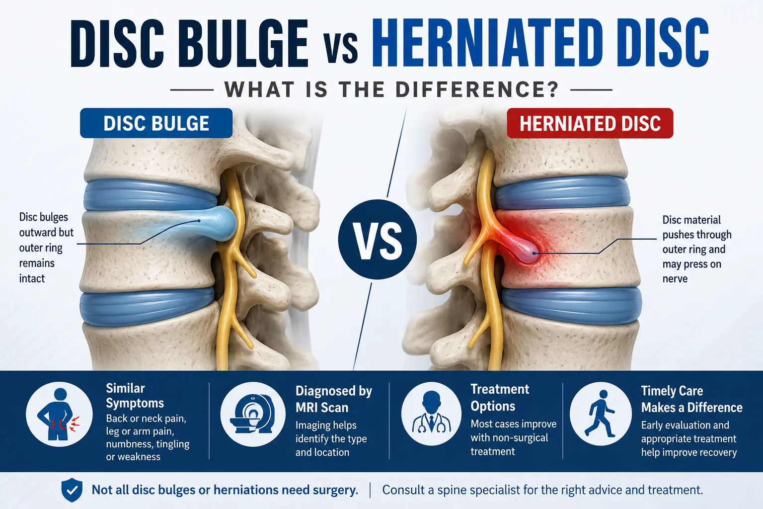
The main disc bulge vs herniated disc difference is the pattern in which disc tissue extends beyond its usual boundary. A disc bulge is generally broader, while a herniated disc is a more localised displacement of disc material. Either finding may cause symptoms if nearby nerves are irritated or compressed, but both can also appear on scans without causing pain.
Understanding the MRI wording is useful, but the scan alone does not decide how serious the problem is or whether treatment is needed.
Disc Bulge vs Herniated Disc: What Is the Main Difference?
A disc bulge generally involves a broader outward extension of the disc, whereas a herniated disc is a more localised displacement of disc material beyond its normal boundary. A herniated disc may be described more specifically as a protrusion or extrusion. Neither finding automatically means that surgery is required.
Here is a simple comparison:
Feature | Disc Bulge | Herniated Disc |
Pattern | Broader extension of the disc | More localised displacement |
Outer disc layer | Usually remains more contained | Disc material extends through a weakened or disrupted area |
MRI terminology | Often reported as a bulge | May be reported as protrusion or extrusion |
Nerve irritation | Possible | Possible and sometimes more likely |
Symptoms | May be absent or cause pain/nerve symptoms | May be absent or cause pain, numbness, tingling or weakness |
Surgery required? | Not simply because a bulge is present | Not simply because a herniation is present |
Standardised lumbar disc terminology distinguishes a generalised bulge from a disc herniation and classifies protrusion and extrusion as patterns of herniation. These imaging descriptions do not, by themselves, establish the cause of a patient’s symptoms or determine treatment.
What Happens to a Spinal Disc?
The spine is made up of bones called vertebrae. Between these bones are intervertebral discs that help absorb forces and allow the spine to move.
Each disc has two main parts:
- A softer central portion called the nucleus pulposus
- A stronger outer ring called the annulus fibrosus
With age and repeated mechanical stress, discs may gradually lose water content and flexibility. The outer portion may extend beyond its normal margin, producing a spinal disc bulge.
In a herniated disc, disc material becomes displaced through a weakened or disrupted part of the outer disc. A herniation may remain relatively contained as a protrusion or extend farther as an extrusion.
This distinction explains why terms such as disc protrusion vs herniation are sometimes confusing. A protrusion is not necessarily a completely separate condition; in standard imaging terminology, it is a type of disc herniation.
Is a Herniated Disc More Serious Than a Bulging Disc?
Not necessarily.
A herniated disc can be more likely to irritate a nearby nerve because the displaced material may extend farther into the space around a nerve root. However, the appearance of the MRI does not always match the severity of symptoms.
A person may have a sizeable disc abnormality with little discomfort, while another person with a smaller abnormality may experience significant leg or arm pain if it affects a nerve in a sensitive location.
Both bulging and herniated discs can also be found incidentally on imaging performed for another reason.
This is why doctors consider three things together:
- The patient’s symptoms
- Findings from the neurological examination
- The location and appearance of the abnormality on imaging
An MRI finding should not be treated simply because it appears abnormal on the report.
Do Disc Bulges and Herniated Discs Cause Different Symptoms?
The symptoms can overlap considerably.
What a person feels often depends more on which nerve is affected and where the disc problem is located than on whether the MRI uses the word “bulge” or “herniation.”
Symptoms in the lower back
A lumbar disc problem may cause:
- Lower back pain
- Pain spreading into the buttock or leg
- Sciatica-like shooting or burning pain
- Tingling or pins and needles
- Numbness in part of the leg or foot
- Weakness in the leg or foot
Symptoms in the neck
A cervical disc problem may cause:
- Neck pain
- Pain around the shoulder blade
- Pain travelling down an arm
- Tingling or numbness in the hand or fingers
- Weakness in the arm, hand or grip
Herniated discs affecting a nerve root can produce radiating pain, numbness, tingling or weakness along the area supplied by that nerve.
Back or neck pain can also arise from muscles, joints, ligaments and other spinal conditions. Symptoms therefore need to be assessed rather than assumed to come from a disc seen on MRI.
Bulging Disc vs Slipped Disc: Are They the Same?
No. The phrase bulging disc vs slipped disc can be confusing because “slipped disc” is an informal term rather than a description of a disc physically slipping out of position.
A slipped disc usually refers to a herniated disc. Part of the disc material becomes displaced beyond its normal boundary; the entire disc does not slide out between the vertebrae.
A bulging disc, in comparison, describes a broader extension of the disc.
Your MRI report may use more precise terms, including:
- Disc bulge
- Disc protrusion
- Disc extrusion
- Disc herniation
- Nerve root compression
- Spinal canal or foraminal narrowing
The clinical importance of each finding depends on its location and whether it corresponds with the patient’s symptoms.
How Is a Disc Problem Diagnosed?
Diagnosis usually begins with a detailed discussion of the symptoms and a physical examination.
A doctor may ask:
- Where did the pain begin?
- Does it travel into an arm or leg?
- Is there numbness or tingling?
- Has muscle strength changed?
- What movements make the symptoms worse?
- Did the symptoms begin after lifting, twisting or another activity?
A neurological examination may include checking muscle strength, sensation, reflexes and walking.
MRI scan
MRI, or magnetic resonance imaging, provides detailed images of the spinal discs, nerve roots, spinal canal and surrounding structures. It can help show whether a disc is bulging or herniated and whether nearby neural structures are affected.
X-rays cannot show a disc herniation directly but may be useful in selected cases to assess bones, alignment or other possible causes of symptoms.
The aim is not simply to identify an abnormal disc. The important question is whether the imaging finding explains the patient’s symptoms.
How Are Disc Bulges and Herniated Discs Treated?
Treatment is based on symptoms and neurological findings rather than the MRI label alone.
Many patients can initially be managed without surgery, depending on their clinical condition.
Non-surgical treatment
A doctor may recommend a combination of:
- Temporary modification of activities that worsen symptoms
- Appropriate pain-relieving or anti-inflammatory medicines
- Guided physiotherapy
- Exercises to improve mobility and spinal support
- Gradual return to normal activities
- Selected spinal injections when clinically appropriate
Long periods of complete bed rest are generally not the goal. Movement and rehabilitation are usually introduced according to pain levels and medical advice.
For patients with persistent symptoms or nerve-related problems, a specialist assessment for slip disc treatment can help determine whether continued conservative care, further investigation or another treatment approach is appropriate.
Does a Herniated Disc Always Need Surgery?
No. A herniated disc does not automatically require an operation. Many people improve with non-surgical management. Surgery is generally considered when symptoms and examination findings justify it rather than simply because a herniation appears on an MRI.
Surgical evaluation may become appropriate when there is:
- Persistent disabling nerve pain despite suitable treatment
- Progressive muscle weakness
- Significant nerve compression that matches the symptoms
- Difficulty walking because of neurological weakness
- Certain urgent neurological problems
If surgery is considered, the exact procedure depends on the level and type of compression, the patient’s symptoms, general health and individual scan findings.
Which Warning Signs Need Urgent Medical Attention?
Most disc-related back pain is not an emergency. Certain neurological symptoms, however, should not be ignored.
Seek urgent medical assessment if back pain or leg symptoms are accompanied by:
- New loss of bladder or bowel control
- Inability or increasing difficulty passing urine
- New numbness around the inner thighs, buttocks or genital area
- Rapidly developing or worsening leg weakness
- Major difficulty standing or walking because of weakness
Bladder or bowel dysfunction together with numbness in the saddle area can occur with cauda equina syndrome, a rare but serious compression of the nerves at the lower end of the spinal canal that requires prompt medical assessment.
When Should You Consult a Neurosurgeon?
A neurosurgical consultation does not automatically mean that surgery will be advised.
Specialist review may be useful when pain continues despite appropriate treatment, symptoms repeatedly return, there is significant arm or leg pain, numbness is persistent, muscle weakness develops, or the MRI shows nerve compression that needs clinical interpretation.
Patients with spinal disc conditions may be evaluated by a neurosurgeon such as Dr. Mazda K. Turel, who can review the history, neurological examination and imaging together before discussing suitable treatment options.
The goal is to understand whether the disc finding is actually responsible for the symptoms and what level of treatment is justified.
Conclusion
Understanding disc bulge vs herniated disc starts with recognising that these terms describe different patterns of disc change, not automatically different levels of danger. A disc bulge is generally broader, while a herniated disc represents a more localised displacement that may be described as a protrusion or extrusion.
More importantly, an MRI report should never be interpreted in isolation. Some disc abnormalities cause significant nerve symptoms, while others are incidental findings that require no specific treatment.
Persistent radiating pain, numbness, weakness or difficulty with normal activities deserves medical evaluation. A specialist can match the symptoms with examination and imaging findings and determine whether rehabilitation, medication, injections or surgical evaluation is appropriate for the individual situation.
Frequently Asked Questions
1. What is the easiest way to understand the disc bulge vs herniated disc difference?
A disc bulge is generally a broader extension of the disc beyond its usual boundary. A herniated disc is a more localised displacement of disc material and may be described as a protrusion or extrusion. Either can be painless or symptomatic. The location of the abnormality and its effect on nearby nerves are often more clinically important than the wording alone.
2. Is a disc protrusion the same as a herniated disc?
A disc protrusion is generally considered a form of disc herniation in standard lumbar MRI terminology. In a protrusion, the displaced portion remains relatively contained compared with an extrusion. Therefore, in a disc protrusion vs herniation comparison, “herniation” is the broader category and “protrusion” describes one particular shape or pattern of that herniation.
3. Can a spinal disc bulge heal without surgery?
Many people with a spinal disc bulge can be managed without surgery, particularly when there is no progressive neurological weakness or other concerning finding. Treatment may involve activity adjustment, appropriate medication and physiotherapy. The need for treatment depends on symptoms rather than the scan alone, because some disc bulges produce no symptoms at all.
4. Which is more painful, a bulging disc or a herniated disc?
A herniated disc may be more likely to cause nerve-related pain because displaced disc material can irritate or compress a nerve root. However, this is not a fixed rule. A bulge positioned against a nerve may be symptomatic, while a herniation elsewhere may cause no pain. Clinical examination is needed to understand the source of symptoms.
5. Can an MRI tell whether my back pain is caused by a disc?
An MRI can identify disc abnormalities and show whether a disc is close to or compressing a nerve. It cannot always prove that the abnormality is the source of pain. Doctors compare the MRI level and side with the patient’s symptoms, strength, sensation and reflexes before deciding whether the finding is clinically relevant.
6. Does “slipped disc” mean the disc has moved out of place?
No. The entire disc does not usually slip out from between the vertebrae. “Slipped disc” is a commonly used term for a herniated disc, in which part of the disc material extends beyond its usual boundary. Medical reports generally use more precise descriptions such as herniation, protrusion or extrusion.
7. When does a herniated disc become an emergency?
Urgent assessment is needed if symptoms include new bladder or bowel problems, difficulty passing urine, numbness around the genital or saddle area, or rapidly worsening leg weakness. These symptoms can indicate significant nerve compression. Severe pain alone does not necessarily mean there is an emergency, but new neurological changes should be assessed promptly.