What Does a Brain Tumor Look Like on MRI?
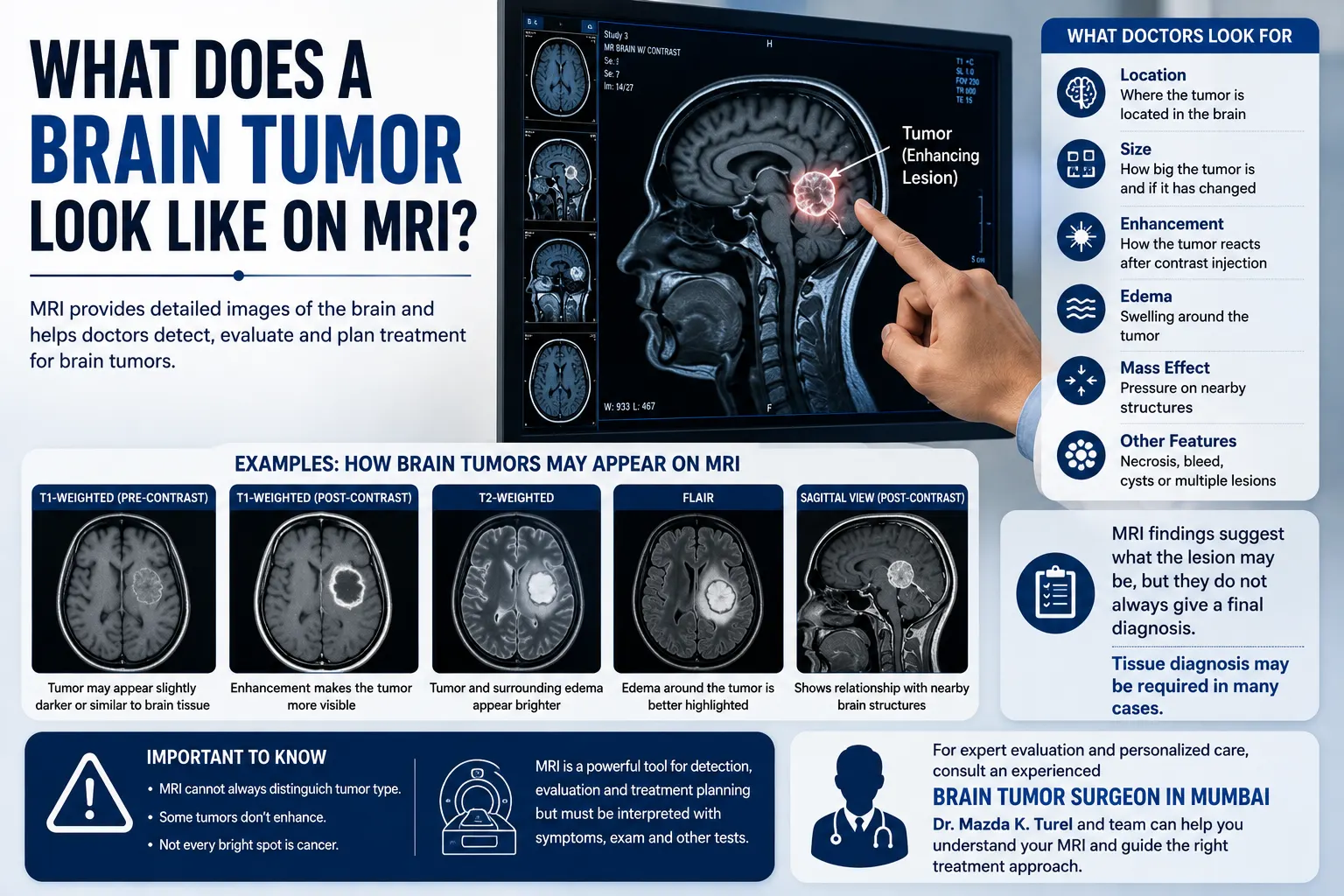
Seeing words such as “lesion,” “mass,” “enhancement,” or “edema” on an MRI report can understandably raise questions. A brain tumor on MRI does not have one fixed appearance. Depending on the type of tumor and the MRI sequence being viewed, it may appear brighter or darker than surrounding brain tissue, enhance after contrast injection, cause swelling, or push nearby structures out of their normal position.
MRI is one of the main imaging tools used to investigate suspected brain tumors because it provides detailed pictures of the brain and surrounding soft tissues without using ionising radiation.
However, MRI appearance alone does not always reveal exactly what a lesion is. Doctors interpret the scan together with symptoms, examination findings, previous imaging and, when required, tissue diagnosis.
What Does a Brain Tumor Look Like on MRI?
A brain tumor may appear on MRI as an abnormal area that differs from normal brain tissue in its signal, shape, borders or response to contrast. Doctors also look for surrounding swelling, pressure on nearby structures, cystic or necrotic areas, bleeding and changes between current and previous scans. No single MRI appearance identifies every brain tumor.
MRI produces several sets of images known as sequences. Each sequence highlights tissue differently. This means the same tumor may look relatively dark on one image and bright on another.
Rather than judging one picture, a radiologist looks across the complete MRI study.
Some features that may be described include:
- Location of the abnormality
- Size and shape
- Whether its margins appear well defined or less distinct
- Signal characteristics on different MRI sequences
- Contrast enhancement
- Swelling around the lesion
- Pressure on nearby brain structures
- Whether the lesion contains fluid, blood or areas of tissue breakdown
- Whether there are one or several lesions
- Changes compared with previous scans
MRI has high sensitivity for detecting many brain abnormalities and assessing the surrounding tissues.
Why Does the Same Tumor Look Different on Different MRI Images?
A brain MRI tumor is not evaluated from a single photograph. MRI changes the way it measures signals from tissues to create different types of images.
Common sequences include T1-weighted, T2-weighted and FLAIR images. Radiologists may also use diffusion-sensitive sequences and post-contrast images.
For patients, the easiest way to understand this is to think of each sequence as showing the brain through a different filter. One may help show anatomy clearly, while another makes fluid or swelling more visible.
This is why trying to identify a tumor by looking at one screenshot from an MRI can be misleading.
What Does “Enhancement” Mean on a Brain MRI?
Contrast-enhanced MRI may be performed when doctors need additional information about a suspected brain abnormality.
The contrast material commonly used for MRI contains gadolinium. It is injected into a vein and can make certain tissues or abnormalities appear more prominent on post-contrast images.
When an MRI report says that a lesion enhances, it means that the area has taken up contrast differently from surrounding tissue.
Enhancement can appear in different patterns, including:
- Relatively uniform enhancement
- Irregular enhancement
- Nodular enhancement
- Enhancement around the edge of a lesion
- Little or no visible enhancement
For example, some metastatic deposits in the brain can show enhancement around their periphery, sometimes described as ring enhancement. However, ring enhancement is not unique to metastatic cancer and should never be interpreted without the full clinical and imaging context.
An enhancing lesion does not automatically mean cancer.
Inflammation, infection and other neurological conditions can also enhance.
What Are Doctors Looking for in MRI Findings of a Brain Tumor?
The wording in a radiology report can sound technical, but many terms describe how the abnormality is affecting the surrounding brain.
Location
Location is particularly important in neurosurgery.
A relatively small tumor in an area involved in speech, movement, vision or other essential functions may have different clinical implications from a larger lesion elsewhere.
The scan helps doctors understand which brain structures are close to the tumor and whether symptoms could reasonably relate to its position.
Size
The radiologist usually measures a lesion in more than one dimension.
If previous scans are available, doctors can compare them to determine whether the abnormality is stable, smaller or has increased in size. Comparing current and previous imaging can significantly help interpretation and follow-up decisions.
Surrounding Edema
Edema means swelling caused by increased fluid within brain tissue.
An MRI may show swelling around a tumor, sometimes extending beyond the obvious borders of the mass. Swelling can contribute to neurological symptoms and may affect treatment planning.
MRI can show edema well, although imaging does not always perfectly distinguish tumor tissue from surrounding fluid-related changes.
Mass Effect
The term mass effect means that a lesion or associated swelling is pushing on nearby brain structures.
Depending on its severity and location, this can include compression of nearby brain tissue or fluid spaces.
A report mentioning mass effect does not by itself state what type of lesion is present. It describes the physical effect the abnormality is having inside the skull.
Midline Shift
The brain has a natural central line separating its two hemispheres.
A sufficiently large mass or substantial swelling can push structures away from this normal position. Radiologists may describe this as midline shift.
The amount of shift is considered along with symptoms, neurological findings and the remainder of the scan.
Cystic or Necrotic Areas
Some lesions contain areas that look different from solid tumor tissue.
A report may describe a cystic component, meaning an area containing fluid, or necrosis, referring to areas of tissue breakdown.
These findings can help narrow the possibilities, but they usually cannot provide a final diagnosis by themselves.
Does Every Brain Lesion on MRI Mean a Tumor?
No.
A brain lesion MRI report simply indicates that an area looks different from expected brain tissue. “Lesion” is a broad imaging term rather than a diagnosis.
Abnormal areas on MRI may be related to several conditions, such as:
- Tumors
- Stroke
- Infection
- Inflammation
- Demyelinating disease
- Previous injury
- Vascular abnormalities
- Other structural changes
Brain MRI is used for many conditions besides tumors, including stroke, epilepsy, multiple sclerosis, infection, traumatic injury and hydrocephalus.
This is why the word “lesion” on an MRI report should not automatically be interpreted as cancer.
Can MRI Tell Whether a Brain Tumor Is Benign or Malignant?
MRI can provide important clues, but it cannot reliably determine the exact diagnosis in every case.
Certain imaging characteristics may make doctors more suspicious of one tumor type than another. The radiologist may consider factors such as:
- Tumor borders
- Pattern of contrast enhancement
- Surrounding edema
- Growth between scans
- Diffusion characteristics
- Presence of necrosis
- Number and distribution of lesions
- Relationship with nearby structures
Imaging characteristics can help predict the nature of some tumors, but radiological patterns can sometimes be misleading. NCI guidance notes that biopsy confirmation is important for many suspected primary brain tumors, although selected lesions with convincing benign clinical and radiological features may sometimes be monitored without biopsy.
Therefore, terms such as “suggestive of” or “likely” in an MRI report should not be confused with a final tissue diagnosis.
Why Might Doctors Order MRI With and Without Contrast?
A doctor may request both non-contrast and contrast-enhanced images because the combination can provide more information about an abnormal area.
The non-contrast sequences show the brain using several different tissue characteristics. After contrast is given, doctors can examine whether the lesion enhances and in what pattern.
Brain MRI may be performed either with or without intravenous contrast depending on the symptoms and the clinical question being investigated.
Contrast is not appropriate in every situation. Before contrast administration, the healthcare team considers factors such as medical history and kidney-related concerns where relevant.
Patients should also tell the MRI team about implants, medical devices, previous operations and pregnancy before entering the scanner because MRI uses a powerful magnetic field.
What Happens After a Brain Tumor Is Seen on a Scan?
Finding a mass on a brain tumor scan is the beginning of the diagnostic process, not necessarily the end.
The next step depends on what the imaging shows.
Doctors may recommend:
- Comparison with previous scans
- A more detailed MRI
- MRI with contrast if not already performed
- Follow-up imaging after a defined interval
- Additional specialised imaging in selected cases
- Evaluation by a neurologist, neurosurgeon or oncologist
- Biopsy
- Surgical removal when appropriate
Radiology reports may recommend follow-up imaging, comparison with earlier scans, clinical correlation or biopsy depending on the finding.
If tissue is obtained, a pathologist examines it to determine the tumor type and other characteristics that can affect treatment planning. NCI guidance notes that biopsy or tissue obtained during surgery is commonly used to establish the diagnosis of a brain tumor.
How MRI Findings Influence Treatment Planning
MRI does more than detect a tumor. It can help the treating team understand whether and how an operation might be approached.
The scan may help assess:
- Exact tumor location
- Approximate extent of the lesion
- Relationship with important brain structures
- Associated swelling
- Pressure on surrounding structures
- Whether multiple abnormalities are present
- Whether the tumor appears to have changed over time
Treatment is then individualised according to the suspected or confirmed diagnosis, symptoms, age, general health, scan findings and potential risks of intervention.
Depending on the condition, management may include observation, surgery, radiation therapy, chemotherapy, targeted treatment or a combination of approaches.
Not every tumor seen on MRI requires immediate surgery.
When Should You Consult a Neurosurgeon?
A neurosurgical review may be appropriate when an MRI identifies a brain mass or lesion that might require biopsy, surgery, monitoring or further assessment.
The neurosurgeon does not look only at the written report. Reviewing the actual MRI images is important because treatment decisions can depend on the precise location of the abnormality and its relationship with surrounding brain structures.
A consultation may cover:
- What the MRI finding appears to represent
- Whether more imaging is necessary
- Whether previous scans should be compared
- Whether observation is reasonable
- Whether tissue diagnosis is required
- Whether surgery has a role
- Potential neurological risks
- Alternative treatment approaches
Patients with an identified brain lesion can seek evaluation from a brain tumor surgeon in Mumbai to review the imaging and understand the possible next steps.
Dr. Mazda K. Turel can assess the MRI alongside the patient’s symptoms, neurological examination and available medical records before discussing whether monitoring, additional testing, surgery or another form of treatment may be appropriate.
Conclusion
A brain tumor on MRI can appear in many ways. Doctors do not diagnose it simply by looking for one bright or dark spot. They assess the lesion’s location, signal on multiple MRI sequences, contrast enhancement, surrounding edema, mass effect, borders and changes over time.
MRI can provide strong clues about what an abnormality may represent and is extremely useful for treatment planning, but imaging does not always establish the exact tumor type. In many situations, biopsy or tissue obtained during surgery is needed for a definitive diagnosis.
If an MRI report describes a brain mass or uncertain lesion, the most useful next step is to have the actual images reviewed alongside the symptoms and neurological findings. This allows decisions about observation, further investigation or treatment to be based on the complete clinical picture.
Frequently Asked Questions
What colour is a brain tumor on MRI?
A brain tumor does not have one standard “colour” on MRI. MRI images are usually displayed in shades of grey, and the same tumor may appear brighter or darker depending on the sequence being viewed. Contrast can make certain lesions appear more prominent. Radiologists interpret several sequences together rather than judging the tumor from its colour on one image.
Does a bright spot on MRI mean a brain tumor?
No. A bright area may represent many different tissue changes depending on the MRI sequence. Stroke, inflammation, demyelination, infection, age-related changes and other conditions can all create abnormal MRI signals. A radiologist interprets the location, pattern, sequences and clinical history before deciding what the finding may represent.
Can an MRI confirm brain cancer?
MRI can strongly suggest that a tumor is present and provide valuable information about its location and characteristics, but it does not always establish the exact tumor type. For many suspected tumors, tissue examination through biopsy or surgery is required for definitive diagnosis and classification.
What does enhancement mean in MRI findings of a brain tumor?
Enhancement means an area becomes more visible after MRI contrast material is administered because it takes up contrast differently from surrounding tissues. The pattern of enhancement can provide diagnostic clues, but enhancement is not exclusive to cancer. Other conditions, including inflammation and infection, can also enhance.
What is the difference between a brain lesion and a brain tumor?
A brain tumor is an abnormal growth of cells. A brain lesion is a broader term for any area that appears abnormal on imaging. A lesion may represent a tumor, but it can also result from stroke, infection, inflammation, injury or another neurological condition. Further assessment is often needed to determine the diagnosis.
Can a small brain tumor be seen on MRI?
MRI can detect many relatively small brain abnormalities because it provides detailed soft-tissue images. However, detection depends on the lesion’s location, characteristics, MRI technique, image quality and whether contrast or specialised sequences are required. MRI is widely used for detecting and evaluating brain tumors and their surrounding tissues.
Why does my doctor want to compare my new MRI with an older scan?
Comparison shows whether a lesion has appeared, changed in size or remained stable. Stability over time can be clinically meaningful, while interval growth may alter the diagnostic possibilities or treatment plan. Radiologists specifically review previous relevant imaging whenever available because it can affect their interpretation and recommendations.