Brain Tumor MRI With Contrast: Why Is Contrast Used?
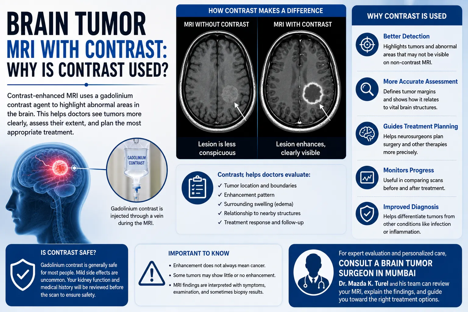
Being told that an MRI needs “contrast” can raise a new set of questions, especially when the scan is being performed for a possible brain tumor. A brain tumor MRI with contrast uses an intravenous contrast agent, usually containing gadolinium, to make certain abnormalities easier to distinguish from surrounding brain tissue.
Contrast can help doctors study a lesion’s borders, enhancement pattern, relationship with nearby structures, and changes following treatment. However, enhancement is only one part of MRI interpretation. A lesion that enhances is not automatically cancerous, and some brain tumors may show little or no enhancement.
Doctors therefore interpret contrast images alongside non-contrast MRI sequences, symptoms, neurological examination findings, previous scans, and sometimes tissue analysis before reaching a diagnosis or recommending treatment.
Why Is Contrast Used for a Brain Tumor MRI?
Contrast is used during brain MRI to make certain tissues and abnormalities more visible. Gadolinium-based contrast changes the MRI signal in areas where it reaches tissues differently, helping radiologists identify patterns of enhancement. This can provide useful information about a suspected tumor’s location, extent, borders, and relationship with surrounding brain structures.
An MRI already produces detailed images without contrast. The contrast portion adds another layer of information rather than replacing the standard scan.
Depending on the clinical situation, a doctor may request:
- MRI without contrast
- MRI with contrast
- MRI both before and after contrast administration
For suspected central nervous system tumors, contrast-enhanced MRI can help demonstrate tumor enhancement and associated findings such as swelling around the lesion.
What Is Gadolinium in a Brain MRI?
Gadolinium brain MRI refers to an MRI examination in which a gadolinium-based contrast agent is given, usually through a small intravenous line in the arm.
Gadolinium-containing contrast material changes the magnetic properties of nearby water molecules, which alters how certain tissues appear on MRI. This helps radiologists see differences that may not be as obvious on non-contrast images.
MRI contrast is different from the iodine-based contrast commonly used for CT scans.
It is also not radioactive. MRI itself uses a strong magnetic field, radio waves, and computer processing rather than ionising radiation.
What Does “Enhancement” Mean on a Contrast MRI?
This is one of the most common words patients notice in an MRI report.
Enhancement means that an area looks more prominent after contrast is administered because it takes up or interacts with the contrast differently from surrounding tissue.
In the brain, the blood-brain barrier normally limits the movement of many substances from blood vessels into brain tissue. Certain tumors and other disease processes can alter this barrier, making contrast-related signal changes more visible.
A radiologist may describe enhancement as:
- Homogeneous or relatively uniform
- Heterogeneous or uneven
- Nodular
- Linear
- Peripheral
- Ring-like
- Minimal
- Absent
These patterns can provide clues, but enhancement alone does not establish a final diagnosis.
Does an Enhancing Lesion Mean Cancer?
No.
An enhancing brain lesion MRI can have several causes. Tumors are one possibility, but infection, inflammation, demyelinating conditions, vascular abnormalities, and other neurological processes may also enhance.
Likewise, the absence of strong enhancement does not rule out a tumor.
This distinction matters because patients sometimes read “enhancing lesion” on a report and assume it means malignant cancer. In reality, the entire MRI appearance and clinical context have to be considered.
What Do Doctors Look for on a Contrast MRI Brain Tumor Scan?
A contrast MRI brain tumor assessment involves much more than checking whether an abnormality becomes brighter after the injection.
Radiologists and treating specialists may examine several features.
Tumor Location
Location often has major clinical importance.
Doctors assess whether the lesion is close to areas responsible for:
- Movement
- Speech
- Vision
- Memory
- Sensation
- Balance
- Hormonal function
- Other essential neurological functions
For a neurosurgeon, understanding the relationship between the tumor and important brain structures is particularly relevant when considering whether surgery is feasible.
Size and Extent
The tumor is generally measured in several dimensions.
Doctors also assess whether an abnormal MRI signal extends beyond the main enhancing portion of the lesion.
If previous scans exist, comparison helps establish whether the abnormality is stable, shrinking, or increasing in size.
Enhancement Pattern
The way a lesion enhances can contribute to the differential diagnosis—the list of conditions doctors think might explain the finding.
However, MRI findings do not always reveal the exact tumor type. Tissue diagnosis may still be necessary in many cases.
Edema
Edema means swelling caused by excess fluid within brain tissue.
MRI can show swelling surrounding some tumors. This may contribute to symptoms and can create additional pressure within the skull.
Mass Effect
Mass effect describes pressure produced by the lesion or associated swelling on nearby brain structures.
A scan may show compression of surrounding tissue or the fluid-filled spaces of the brain.
Midline Shift
If a mass or swelling pushes central brain structures away from their normal position, the radiologist may describe midline shift.
This finding is interpreted together with the size of the lesion, swelling, symptoms, and neurological examination.
Is MRI With Contrast Better Than MRI Without Contrast?
Neither scan should simply be thought of as “better.”
They provide different information.
Non-contrast MRI sequences can reveal anatomy, swelling, fluid changes, bleeding, diffusion characteristics, and many other features. Contrast-enhanced images show whether and how an abnormality enhances.
A brain tumor MRI with contrast often includes both sets of images so that doctors can compare the appearance before and after gadolinium is administered. Brain MRI may be ordered with or without intravenous contrast depending on the symptoms and what the healthcare team is investigating.
For this reason, a patient should not assume that a scan performed without contrast is incomplete. The appropriate imaging protocol depends on the clinical question.
Can Contrast Tell Doctors What Type of Brain Tumor It Is?
Not always.
Contrast enhancement may help doctors narrow the possible diagnoses, but there is no enhancement pattern that identifies every brain tumor with certainty.
MRI findings may suggest a particular type of lesion based on:
- Location
- Shape
- Borders
- Enhancement
- Swelling
- Diffusion characteristics
- Presence of bleeding or necrosis
- Number of lesions
- Changes on previous imaging
Even with detailed brain tumor imaging, some abnormalities cannot be classified confidently from MRI alone.
Depending on the suspected diagnosis, doctors may recommend additional imaging, monitoring, biopsy, or surgery to obtain tissue. NCI guidance notes that brain tumors must also be differentiated from other abnormalities that can have similar imaging and clinical appearances.
Is Gadolinium Contrast Safe?
Gadolinium-based contrast agents are widely used, and most patients tolerate them without significant problems when appropriate safety guidelines are followed. Mild reactions can occur, while serious allergic reactions are rare.
Before a contrast MRI, tell the healthcare team if you have:
- Significant kidney disease
- Previous reactions to MRI contrast
- Recent serious illness or surgery
- Pregnancy or possible pregnancy
- Implanted medical devices or metal in the body
Kidney function deserves particular attention in patients with serious kidney disease. A rare condition called nephrogenic systemic fibrosis has been associated with gadolinium exposure, mainly in people with severe kidney problems; it is exceptionally rare with newer agents.
Very small amounts of gadolinium may also remain in the body following some contrast examinations. Current imaging decisions therefore consider whether contrast is clinically necessary, especially in people expected to undergo repeated scans.
Gadolinium contrast is generally avoided during pregnancy unless there is a specific clinical reason and the expected benefit justifies its use.
What Happens During a Brain MRI With Contrast?
The exact process can vary between imaging centres, but it usually follows a straightforward sequence.
You will be asked to remove metal items and confirm information about medical devices, previous procedures, kidney problems, pregnancy, and relevant allergies.
During the examination:
- You lie on the MRI table with your head positioned securely.
- Several non-contrast images may be taken first.
- Gadolinium contrast is administered through an IV when required.
- Additional images are then obtained.
- A radiologist later reviews all sequences together.
MRI scanners make loud knocking or tapping sounds, so hearing protection is normally provided.
Remaining still is important because movement can reduce image quality.
How Does Contrast MRI Help With Brain Tumor Treatment Planning?
Finding a lesion is only one purpose of MRI.
For a diagnosed or suspected brain tumor, the images may help doctors evaluate:
- Where the tumor is located
- How large it is
- How close it is to important structures
- Whether there is surrounding swelling
- Whether there is significant mass effect
- Which areas enhance
- Whether multiple lesions are present
- Whether the appearance has changed over time
These details can influence decisions about observation, biopsy, surgery, radiation therapy, systemic treatment, or further diagnostic testing.
MRI can also be used during follow-up to assess how an abnormality changes over time or after treatment.
MRI findings must still be interpreted alongside the diagnosis, age, symptoms, general health, neurological examination, and individual clinical circumstances.
When Should You Consult a Neurosurgeon?
A neurosurgical review may be appropriate when an MRI identifies a brain mass or lesion that might require biopsy, surgery, monitoring or further assessment.
The neurosurgeon does not look only at the written report. Reviewing the actual MRI images is important because treatment decisions can depend on the precise location of the abnormality and its relationship with surrounding brain structures.
A consultation may cover:
- What the MRI finding appears to represent
- Whether more imaging is necessary
- Whether previous scans should be compared
- Whether observation is reasonable
- Whether tissue diagnosis is required
- Whether surgery has a role
- Potential neurological risks
- Alternative treatment approaches
Patients with an identified brain lesion can seek evaluation from a brain tumor surgeon in Mumbai to review the imaging and understand the possible next steps.
Dr. Mazda K. Turel can assess the MRI alongside the patient’s symptoms, neurological examination and available medical records before discussing whether monitoring, additional testing, surgery or another form of treatment may be appropriate.
Conclusion
A brain tumor on MRI can appear in many ways. Doctors do not diagnose it simply by looking for one bright or dark spot. They assess the lesion’s location, signal on multiple MRI sequences, contrast enhancement, surrounding edema, mass effect, borders and changes over time.
MRI can provide strong clues about what an abnormality may represent and is extremely useful for treatment planning, but imaging does not always establish the exact tumor type. In many situations, biopsy or tissue obtained during surgery is needed for a definitive diagnosis.
If an MRI report describes a brain mass or uncertain lesion, the most useful next step is to have the actual images reviewed alongside the symptoms and neurological findings. This allows decisions about observation, further investigation or treatment to be based on the complete clinical picture.
Frequently Asked Questions
Why do doctors order a brain tumor MRI with contrast?
A brain tumor MRI with contrast can make certain abnormalities more visible and show how a lesion enhances after gadolinium administration. Doctors use this information alongside non-contrast sequences to assess the lesion’s location, borders, surrounding swelling, and relationship with nearby structures. Contrast can improve diagnostic information, but it does not always identify the exact tumor type.
Does every brain tumor enhance with gadolinium?
No. Brain tumors vary in how much they enhance. Some show marked contrast enhancement, others enhance only in certain areas, and some may demonstrate little or no enhancement. The lack of enhancement does not by itself exclude a tumor. Radiologists assess all MRI sequences and the overall clinical context rather than relying on contrast appearance alone.
Does contrast make a brain tumor look bigger?
Contrast does not physically enlarge the tumor. It can make certain parts of a lesion easier to see, which may change how clearly its enhancing component can be measured. Other MRI sequences may reveal additional non-enhancing abnormalities or surrounding edema, so doctors use the complete study to assess the lesion’s extent.
What does “no abnormal enhancement” mean on a brain MRI?
It means that the radiologist did not identify an area showing unexpected contrast enhancement on the post-contrast images. This can be reassuring in some clinical situations, but it does not mean that every possible neurological disorder or tumor has been excluded. The rest of the MRI findings and the reason for the scan still matter.
Is MRI contrast for a brain lesion always necessary?
No. Whether MRI contrast for a brain lesion is needed depends on the clinical question, previous imaging, symptoms, and what doctors are trying to distinguish. Some brain abnormalities can be evaluated without contrast, while others benefit substantially from post-contrast images. The ordering doctor and radiologist determine the appropriate MRI protocol.
Can gadolinium MRI determine whether a tumor is malignant?
Gadolinium enhancement can provide useful clues but cannot reliably determine malignancy by itself. Benign lesions can enhance, and some tumors may show limited enhancement. Doctors consider multiple MRI features, the clinical history, examination findings, and often tissue diagnosis before determining the exact nature of a brain tumor.
Can I have a contrast MRI if I have kidney disease?
It depends on the severity of the kidney problem and the type of contrast agent being considered. Patients with significant kidney disease should inform their radiology team before the examination. Doctors may review kidney function and weigh the benefit of contrast against potential risk before proceeding.