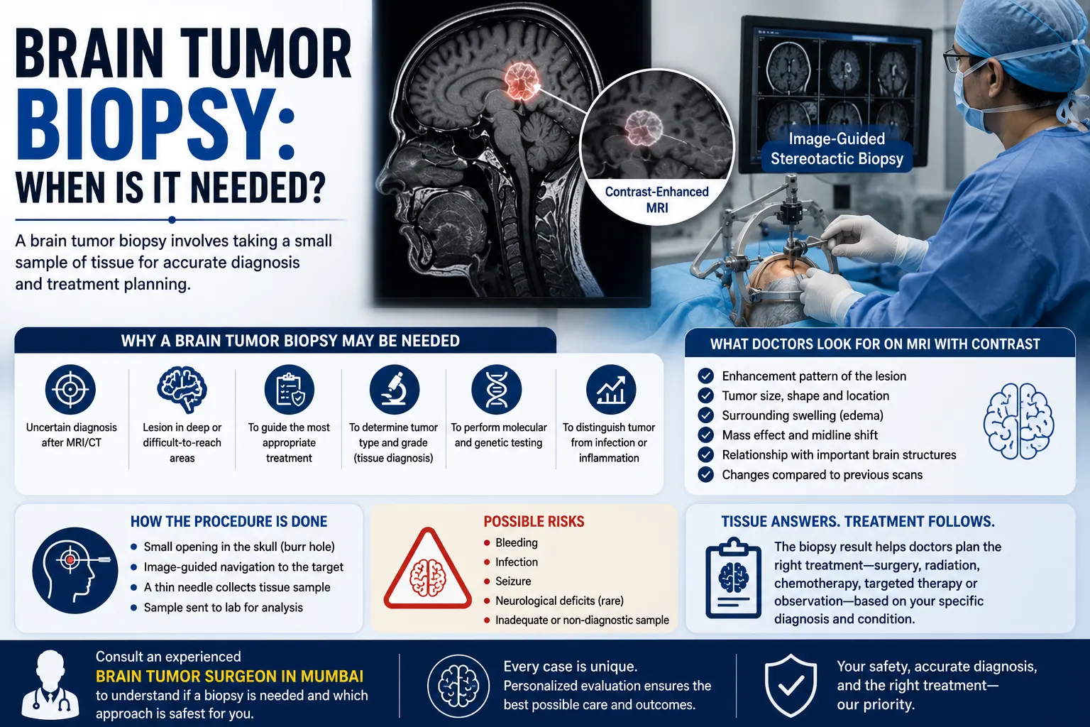
Finding an abnormal area on a brain MRI does not always tell doctors exactly what it is. In some situations, a brain tumor biopsy is needed to obtain a small sample of tissue so that a pathologist can examine it and establish a more precise diagnosis. The tissue may also undergo laboratory and molecular testing that can influence treatment planning.
A biopsy is not automatically necessary for every brain lesion. Some tumors can be diagnosed with a high degree of confidence from their clinical and imaging features, while others may be monitored. In other situations, obtaining tissue is essential before deciding on surgery, radiation, chemotherapy, targeted treatment, or another approach.
The decision depends on what the MRI shows, where the lesion is located, whether it can be safely removed, the patient’s symptoms and overall health, and how much the biopsy result is likely to change treatment.
What Is a Brain Tumor Biopsy?
A brain tumor biopsy is a procedure in which a neurosurgeon obtains a small sample of abnormal brain or tumor tissue for examination in a laboratory. It may be performed through a targeted needle procedure or during an operation to remove the tumor. The tissue helps doctors identify what type of lesion is present and plan treatment.
A pathologist examines the sample under a microscope and may perform additional laboratory tests. Depending on the suspected tumor, the tissue can provide information about cell type and biological or molecular characteristics.
The National Cancer Institute notes that tissue testing can include immunohistochemistry and analysis of genetic or chromosomal changes. These findings can help distinguish one type of brain tumor from another and may influence management.
In simple terms, the scan tells doctors where the abnormality is and what it looks like. A biopsy can help answer what it actually is.
When Is a Brain Biopsy Needed?
A biopsy for brain tumor may be considered when imaging shows an abnormal lesion but doctors cannot establish the diagnosis confidently from MRI or CT findings alone.
Common situations include:
- The MRI appearance could represent more than one condition
- The exact tumor type needs to be established before treatment
- A lesion is deep within the brain and complete removal may carry significant risk
- Surgery to remove the whole lesion is not currently appropriate
- Doctors need to distinguish a tumor from infection or inflammation
- A patient has a suspected metastatic lesion but the diagnosis is uncertain
- Tissue information is needed to guide radiation, drug treatment, or other therapy
- The lesion has changed and doctors need to understand whether it represents tumor or another process
NCI guidance notes that imaging patterns can sometimes be misleading and that biopsy may be needed to distinguish a primary brain tumor from other conditions such as metastatic disease or infection.
A biopsy may also be useful when the lesion is in a location where removing it completely would carry greater neurological risk but a tissue diagnosis is still needed.
Does Every Brain Tumor Need a Biopsy?
No.
There are situations where a biopsy may not be recommended.
For example, imaging and clinical findings may sometimes strongly suggest a benign lesion that can be monitored safely with repeat imaging rather than immediately sampled or removed. NCI guidance recognises that selected tumors with convincing benign clinical and radiological features may be managed with active surveillance without biopsy.
A biopsy may also be avoided when:
- The location makes tissue sampling unusually risky
- The patient’s health makes an invasive procedure unsafe
- Imaging and other clinical information already provide enough information to guide management
- The lesion can be removed surgically and tissue will be obtained during that operation
This is why the answer to when is brain biopsy needed varies from patient to patient.
The key question is not simply, “Can a biopsy be done?” It is, “Will obtaining tissue provide information that meaningfully helps treatment, and can it be obtained with an acceptable level of risk?”
Stereotactic Brain Biopsy: How Does It Work?
A stereotactic biopsy is a targeted procedure used to obtain tissue from a precise location inside the brain.
Stereotactic means that imaging and computer-guided navigation are used to identify three-dimensional coordinates within the brain. MRI or CT images help the neurosurgeon plan a safe route toward the lesion.
During a typical brain biopsy procedure:
- The patient’s MRI or CT is reviewed to identify the target.
- The head is positioned so that the biopsy can be performed accurately.
- A small area of the scalp is prepared.
- A small incision and opening in the skull, called a burr hole, are made.
- A biopsy needle is guided toward the lesion using stereotactic navigation.
- Small tissue samples are collected.
- The samples are sent to pathology for examination.
Modern procedures may use frame-based or frameless navigation depending on the case and surgical system.
AANS describes stereotactic biopsy as particularly useful for obtaining tissue from deep or difficult-to-reach lesions while limiting disruption to surrounding brain tissue.
Is a Biopsy Different From Brain Tumor Removal?
Yes.
A biopsy is primarily performed to obtain enough tissue for diagnosis. It is not designed to remove the entire tumor.
A resection, on the other hand, is an operation in which the surgeon aims to remove part or as much of the tumor as can be done safely. Tissue obtained during resection is also sent for pathological examination.
Sometimes doctors recommend biopsy rather than immediate tumor removal because:
- The lesion is deep
- It lies close to critical neurological structures
- The diagnosis is uncertain
- Treatment may primarily involve medicines or radiation rather than surgical removal
- The patient’s health makes a larger operation less suitable
In other circumstances, if the tumor can be removed safely and surgery is already indicated, a separate biopsy procedure may not be necessary because tissue can be obtained during the operation. NCI describes both stereotactic biopsy and open biopsy/surgical approaches as ways to establish a brain tumor diagnosis.
What Does a Brain Tumor Tissue Diagnosis Tell Doctors?
A brain tumor tissue diagnosis can provide considerably more detail than simply confirming that abnormal cells are present.
Pathologists may evaluate:
- The type of cells involved
- How abnormal the cells appear
- Tumor characteristics under the microscope
- Specific protein markers
- Selected molecular or genetic alterations
Modern brain tumor classification increasingly combines microscopic findings with molecular information.
This matters because two lesions that look similar on MRI can behave differently or require different treatment approaches.
Tissue results may therefore help the treating team decide whether a patient needs:
- Surgery
- Radiation therapy
- Chemotherapy
- Targeted or other systemic treatment
- Observation
- A combination of treatments
The biopsy does not determine treatment by itself. Doctors combine pathology with the patient’s age, symptoms, neurological examination, imaging findings, tumor location, general health, and individual circumstances.
What Are the Possible Risks of a Brain Biopsy?
A brain biopsy is a surgical procedure, so it is not completely risk-free.
Possible complications can include:
- Bleeding within or around the biopsy area
- Infection
- Seizure
- New neurological symptoms
- Reaction to anaesthesia
- Swelling
- An inadequate or non-diagnostic tissue sample
The neurological risk depends partly on where the lesion is located and what structures need to be passed or approached to reach it.
For example, a lesion near pathways responsible for movement, language, vision, or other important functions may require particularly careful planning.
AANS notes that stereotactic biopsy is planned with imaging and navigation to select a safe and accurate path to the target, but bleeding, infection and failure to obtain diagnostic tissue remain recognised risks.
Before recommending the procedure, the neurosurgeon weighs these risks against the value of obtaining a confirmed diagnosis.
What Happens After the Biopsy?
After the procedure, patients are monitored for neurological changes and other complications.
The healthcare team may check:
- Strength in the arms and legs
- Speech
- Level of alertness
- Vision
- Sensation
- Headache or nausea
- The surgical wound
Some patients may be discharged after a relatively short period of observation, while others may remain in hospital overnight or longer depending on their condition, biopsy location, symptoms, and hospital protocol.
Recovery instructions vary. Patients may temporarily be advised to limit strenuous activity and monitor the wound.
The final pathology report usually takes longer than an immediate preliminary assessment because additional staining and molecular tests may be required.
Which Symptoms After Biopsy Need Urgent Attention?
The treating neurosurgical team should provide specific discharge instructions.
Urgent medical assessment may be needed for symptoms such as:
- New or worsening weakness
- New difficulty speaking
- A seizure
- Significant loss of consciousness or unusual drowsiness
- Rapidly worsening confusion
- Severe or increasing headache
- Repeated vomiting
- New significant vision changes
- Bleeding, discharge, or signs of infection around the wound
These symptoms do not necessarily mean that a serious complication has occurred, but they require prompt assessment after an intracranial procedure.
What Happens Once the Biopsy Result Is Available?
The biopsy result is used together with the MRI and clinical findings to build the treatment plan.
Depending on the diagnosis, the next step may be:
- Continued observation
- Further surgery
- Radiation therapy
- Chemotherapy or another drug treatment
- Additional testing
- Referral to neuro-oncology or radiation oncology
- Multidisciplinary discussion
For some tumors, molecular results may influence which therapies are considered.
A brain biopsy should therefore be viewed as part of a larger diagnostic process rather than an isolated procedure.
When Should You Consult a Neurosurgeon?
A neurosurgical assessment is particularly relevant when an MRI or CT scan shows a brain lesion and there is uncertainty about whether it should be monitored, biopsied, or removed.
The neurosurgeon may review:
- The actual MRI images
- Location and size of the lesion
- The possible diagnoses
- Whether tissue confirmation is necessary
- Whether biopsy or surgical removal is more appropriate
- The safest route to obtain tissue
- Possible neurological risks
- Alternatives to biopsy
- How the result may affect treatment
Patients with a suspected brain tumor can seek evaluation from a brain tumor surgeon in Mumbai to understand whether tissue diagnosis is necessary and which surgical approach, if any, may be suitable.
Dr. Mazda K. Turel can review symptoms, neurological findings, scans, and available reports before discussing whether biopsy, resection, observation, or another approach should be considered.
Conclusion
A brain tumor biopsy is considered when doctors need tissue to determine exactly what a brain lesion represents and when that information is likely to influence treatment. It may be especially useful for deep or uncertain lesions where immediate removal is not the safest or most appropriate option.
Not every brain tumor needs a separate biopsy. Some lesions can be monitored, while others are removed surgically and diagnosed from tissue collected during that procedure.
The decision should come from a careful review of the MRI, symptoms, neurological findings, overall health, and the risks and benefits of obtaining tissue. If biopsy is recommended, understanding why it is needed, what information it may provide, and what alternatives exist can help patients approach the decision with clearer expectations.
Frequently Asked Questions
Is a brain tumor biopsy always necessary before treatment?
No. A brain tumor biopsy is often valuable for establishing a tissue diagnosis, but it is not required in every case. Some lesions have clinical and imaging characteristics that allow observation or treatment without biopsy, while others may be removed surgically and diagnosed from the tissue obtained during that operation. The decision depends on location, imaging, symptoms, safety, and how the result would change management.
Is a stereotactic brain biopsy considered major surgery?
It is an invasive neurosurgical procedure but is generally less extensive than an operation intended to remove a large brain tumor. A small opening is made in the skull, and image-guided navigation is used to direct a biopsy needle toward the target. Recovery and risk depend on the patient’s health, lesion location, and individual procedure.
Can MRI tell what a brain tumor is without a biopsy?
MRI can sometimes strongly suggest a particular diagnosis, but imaging cannot identify every brain lesion with certainty. Different tumors, infections, inflammatory conditions, and other abnormalities may sometimes look similar. When the exact diagnosis will affect treatment, tissue examination may be needed to determine what the lesion represents.
How much tissue is taken during a brain biopsy?
A stereotactic biopsy is designed to remove only small tissue samples needed for laboratory analysis rather than the whole lesion. The neurosurgeon determines how many samples are appropriate based on the lesion, imaging, and intraoperative findings. The goal is to obtain enough representative tissue for diagnosis while limiting unnecessary disruption to surrounding brain.
What happens if a brain biopsy does not give a clear diagnosis?
Occasionally, the tissue obtained may be insufficient or may not represent the part of the lesion needed for a definitive diagnosis. In that situation, doctors review the pathology, imaging, symptoms, and procedural details before deciding whether additional testing, repeat biopsy, surgical removal, or close imaging follow-up is appropriate. AANS recognises a non-diagnostic sample as a possible limitation of stereotactic biopsy.
Can a brain tumor biopsy make the tumor spread?
A properly performed stereotactic brain biopsy is used routinely to diagnose intracranial lesions. Concern about “spreading the tumor” should not be used to avoid a biopsy that a neurosurgical and oncology team believes is necessary. The decision is instead based on whether tissue diagnosis is needed, whether sampling can be performed safely, and how the result will affect treatment.
How do doctors decide between biopsy and removing the tumor?
The decision depends on tumor location, suspected diagnosis, size, symptoms, relationship to critical brain structures, general health, and whether safe removal is feasible. If removal is appropriate, tissue can usually be obtained during surgery. For deep, difficult-to-reach, uncertain, or non-resectable lesions, a stereotactic biopsy may provide the diagnosis needed to plan further treatment.