Being advised to undergo a stereotactic brain biopsy does not necessarily mean that doctors already know a brain lesion is cancerous. In many cases, the reason for the procedure is precisely the opposite: imaging has identified an abnormal area, but MRI or CT alone cannot provide a definite diagnosis.
A stereotactic biopsy allows a neurosurgeon to collect a small amount of tissue from a carefully selected area using image-based navigation. A pathologist then examines that tissue to determine whether the lesion represents a tumor, infection, inflammation, or another condition.
Compared with open brain surgery, the procedure uses a much smaller opening and is primarily performed for diagnosis rather than tumor removal.
What Is a Stereotactic Brain Biopsy?
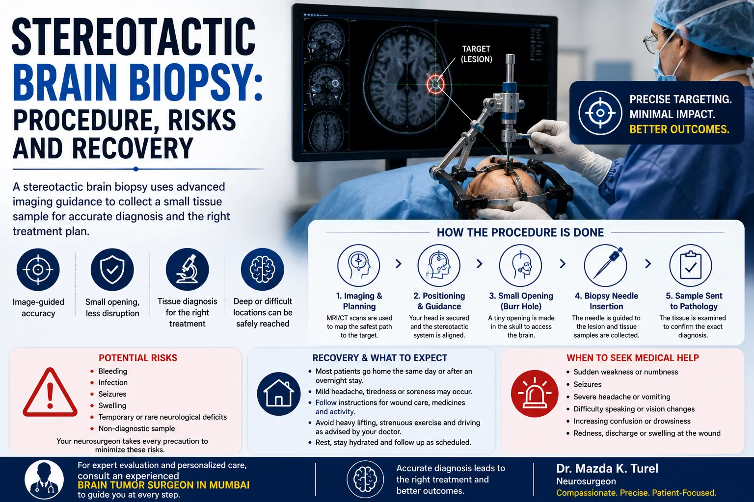
A stereotactic brain biopsy is a neurosurgical procedure that uses MRI or CT images together with computer-guided navigation to direct a biopsy needle to a precise location in the brain. Small tissue samples are removed through a small skull opening and sent to a laboratory for diagnosis.
The word stereotactic refers to the use of three-dimensional imaging and navigation to locate the target accurately.
A stereotactic biopsy brain procedure may be considered when an abnormality is:
- Deep within the brain
- Close to important neurological structures
- Difficult to diagnose from imaging alone
- Not suitable for immediate surgical removal
- Suspected to be a tumor, infection, inflammation, or another disease process
Stereotactic biopsy is often chosen when the primary goal is obtaining a diagnosis while avoiding a larger operation.
Why Might Doctors Recommend a Brain Biopsy?
MRI can provide detailed information about a lesion’s size, location, enhancement, swelling, and relationship with surrounding structures. Yet different diseases can sometimes produce similar-looking scans.
A tissue sample can help answer a question that imaging cannot always answer with certainty: What exactly is this lesion?
Doctors may recommend biopsy to:
- Confirm whether an abnormality is a tumor
- Identify the tumor type
- Distinguish tumor from infection or inflammation
- Investigate an uncertain lesion in a deep or sensitive location
- Assess whether a change after previous tumor treatment represents recurrence or treatment-related changes
- Obtain information needed before deciding on further treatment
A brain biopsy is mainly a diagnostic procedure. It does not usually remove or treat the entire lesion.
How Is an Image Guided Brain Biopsy Planned?
Careful planning begins before the patient enters the operating room.
The neurosurgeon reviews MRI or CT images and identifies both the target and the safest route to reach it. The planned path needs to consider nearby blood vessels and brain regions involved in functions such as movement, speech, sensation, and vision.
The image guided brain biopsy may be performed with either a frame-based or frameless navigation system.
Frameless Stereotactic Biopsy
Many modern procedures use small markers placed on the scalp together with computer navigation. These markers allow the navigation system to match the patient’s head to the MRI or CT images.
The surgeon can then follow the planned trajectory toward the lesion.
Frame-Based Stereotactic Biopsy
In selected cases, a specialised stereotactic frame may be attached to the head. The frame provides fixed reference points that help calculate the exact position of the target.
Both techniques are designed around the same basic principle: reaching a precise area while limiting unnecessary disturbance of surrounding brain tissue.
What Happens Before the Procedure?
Preparation varies according to the patient, hospital, anaesthetic plan, and lesion being investigated.
Your healthcare team will usually review:
- Current medications
- Blood-thinning medicines
- Over-the-counter medicines and supplements
- Previous illnesses and operations
- Seizure history
- Heart or lung conditions
- Diabetes and other long-term medical conditions
- Allergies
- Previous MRI or CT scans
Some medicines that affect bleeding may need to be adjusted before surgery, but patients should not stop prescription medicines on their own. Medication changes should come from the treating team.
Instructions about eating and drinking before anaesthesia will also be provided.
It is sensible to arrange for someone to accompany you home after discharge and to plan for some help during the early recovery period.
How Is a Stereotactic Brain Biopsy Performed?
The details vary, but the procedure generally follows several stages.
1. Positioning and Navigation
The patient’s head is positioned securely. The surgical team then links the patient’s anatomy with the previously obtained MRI or CT images using the navigation system.
This allows the neurosurgeon to confirm the planned target and route.
2. Anaesthesia
A stereotactic biopsy is commonly performed with anaesthesia or medication that keeps the patient comfortable. The exact approach depends on the patient’s condition and the surgical plan.
3. Small Scalp Incision
Only a limited amount of hair may need to be trimmed.
The surgeon makes a small incision in the scalp at the predetermined entry point.
4. Burr Hole
A small opening known as a burr hole is created in the skull.
The protective covering of the brain is then opened sufficiently to allow the biopsy instrument to pass.
5. Biopsy Needle Placement
Using stereotactic navigation, the surgeon guides the needle along the planned path to the target.
Small tissue samples are taken from the lesion.
6. Tissue Examination
The tissue is sent to a pathologist. Depending on the case, an initial assessment may sometimes be performed while the operation is still underway, with more detailed testing completed later.
The final diagnosis may require specialised staining, molecular testing, or additional laboratory analysis.
The needle is then removed and the small incision is closed.
What Are the Main Brain Biopsy Risks?
A stereotactic biopsy uses a smaller approach than open surgery, but it still involves entering the brain and therefore carries genuine surgical risks.
Possible brain biopsy risks include:
Bleeding
Bleeding can occur at or around the biopsy site. Small amounts may cause no symptoms, while more significant bleeding can potentially affect neurological function.
The location of the lesion and nearby blood vessels are therefore considered carefully during planning.
Temporary or Persistent Neurological Changes
Depending on the part of the brain being sampled, possible changes may involve:
- Weakness
- Numbness
- Speech difficulty
- Confusion
- Vision changes
- Coordination problems
The individual risk depends heavily on where the lesion is situated and which structures lie near the planned path.
Seizures
A seizure can occur following a brain biopsy, although this is not expected in every patient.
Patients who already have seizures may also have an individual medication plan around surgery.
Infection
An infection can develop in the scalp, skull, or deeper tissues after any surgical procedure. Sterile surgical technique is used to reduce this risk.
Brain Swelling
Temporary swelling, or edema, can develop around the biopsy area and may contribute to headache, weakness, or confusion.
Non-Diagnostic Sample
Occasionally, the collected tissue does not provide a definite answer. This can happen if the sample is too small or does not adequately represent the abnormal area.
Further imaging, repeat biopsy, or another approach may then be considered.
These risks are why the neurosurgeon evaluates whether obtaining tissue is likely to provide enough clinical benefit to justify the procedure.
What Happens Immediately After the Biopsy?
After surgery, the patient is moved to a recovery area where neurological function and general condition are monitored.
The healthcare team may repeatedly check:
- Alertness
- Speech
- Arm and leg strength
- Sensation
- Vision
- Blood pressure and other observations
- Headache
- Nausea or vomiting
- The incision site
Mild soreness around the incision, fatigue, sleepiness after anaesthesia, or a headache can occur during early recovery.
Some people may be discharged the same day, while others remain in hospital overnight for observation. The decision depends on the patient’s condition, biopsy location, symptoms, anaesthesia, and individual clinical circumstances.
Brain Biopsy Recovery: What Should You Expect at Home?
Brain biopsy recovery is usually shorter than recovery from an open craniotomy because only a small opening is used. However, recovery is individual. Patients should follow their own neurosurgeon’s instructions regarding wound care, exercise, work, driving, and medicines rather than following a fixed timetable.
At home, mild tiredness, scalp tenderness, or a mild headache may occur.
Practical recovery steps may include:
- Take prescribed medicines exactly as directed.
- Keep the incision and dressing clean according to your surgical team’s instructions.
- Rest when tired rather than pushing through significant fatigue.
- Arrange help at home during the early period if needed.
- Avoid scratching or disturbing the wound.
- Attend scheduled follow-up appointments.
- Do not restart medicines that were stopped before surgery unless the treating team advises you to do so.
The surgeon will tell you when the dressing can be removed and when washing the scalp is appropriate.
What About Exercise, Work and Driving?
There is no single recovery schedule that applies to every patient.
Mayo Clinic advises that patients may temporarily need to restrict strenuous exercise, heavy lifting, and other activities after brain biopsy and should receive individual guidance regarding return to work, driving, and exercise.
Your recommendations may depend on:
- Whether seizures have occurred
- Neurological symptoms
- Type of anaesthesia
- Location of the biopsy
- Postoperative imaging
- Medicines being taken
- Type of work you do
- How you feel during recovery
Do not assume that feeling well enough to drive means it is medically or legally appropriate to do so. Ask your treating team specifically when driving can resume.
Which Symptoms After a Brain Biopsy Need Urgent Attention?
Your own discharge instructions should take priority, but certain symptoms deserve prompt medical attention after an intracranial procedure.
Contact your care team urgently or seek emergency medical assessment for:
- New weakness or numbness
- New difficulty speaking
- A new seizure
- Loss of consciousness
- Increasing or severe confusion
- A rapidly worsening headache
- Repeated vomiting
- New significant vision changes
- Increasing difficulty walking
- Unusual or worsening drowsiness
- Significant bleeding or discharge from the incision
- Increasing redness, swelling, or other signs of wound infection
Neurological symptoms after biopsy may sometimes reflect bleeding, swelling, seizure activity, or another complication and should not simply be observed at home without medical advice.
When Will the Biopsy Results Be Available?
A biopsy result is not always available immediately.
The tissue needs to be examined by a pathologist, and additional tests may be required. Depending on the suspected diagnosis, these can include specialised stains and molecular studies.
Mayo Clinic notes that final results may take from several days to longer when detailed analysis is required.
The report may help determine whether the lesion represents:
- A primary brain tumor
- Metastatic cancer
- Infection
- Inflammatory disease
- Another neurological condition
If a tumor is diagnosed, additional tissue characteristics may help guide treatment.
The result is interpreted together with the MRI, neurological examination, symptoms, and other medical information rather than in isolation.
What Happens After the Diagnosis?
The next step depends entirely on what the biopsy finds.
Options may include:
- Observation and repeat imaging
- Surgical removal
- Radiation therapy
- Chemotherapy
- Targeted or other drug treatment
- Treatment for infection or inflammation
- Referral to other specialists
For brain tumors, treatment decisions may involve a multidisciplinary team that includes neurosurgery, radiation oncology, medical or neuro-oncology, neuroradiology, and pathology.
A biopsy is therefore not the end of the process. Its purpose is to provide information that allows the next decision to be made on firmer diagnostic grounds.
When Should You Consult a Neurosurgeon?
A neurosurgical consultation is relevant when brain imaging identifies a lesion and there is a question about whether biopsy is necessary, whether surgical removal would be more appropriate, or whether the abnormality can be safely observed.
A neurosurgeon may review:
- The original MRI or CT images
- Possible diagnoses
- Lesion location and size
- Nearby blood vessels and critical brain structures
- Whether a biopsy will change treatment
- Stereotactic versus open surgical options
- Individual neurological risks
- Recovery expectations
- What happens if the tissue sample is inconclusive
Patients with a suspected brain tumor may seek evaluation from a brain tumor surgeon in Mumbai to discuss whether a stereotactic biopsy or another approach is appropriate.
Dr. Mazda K. Turel can review the patient’s imaging, neurological findings, symptoms, and medical condition before discussing the potential benefits, limitations, and risks of obtaining tissue.
Conclusion
A stereotactic brain biopsy allows doctors to obtain tissue from a precisely targeted brain lesion through a relatively small surgical approach. MRI or CT-based navigation is used to plan the route, while pathology helps determine what the lesion actually represents.
The procedure is generally performed because the information gained from tissue is expected to influence diagnosis or treatment. Like any intracranial procedure, it carries risks, including bleeding, seizures, infection, swelling, and neurological changes.
Brain biopsy recovery is individual. Patients should follow their own instructions for incision care, activity, medicines, driving, and follow-up rather than relying on a fixed timeline.
Understanding why the biopsy is being recommended, what the pathology can clarify, and how the result may change treatment can make the procedure and the next steps easier to approach.
Frequently Asked Questions
Is stereotactic brain biopsy the same as brain surgery?
A stereotactic brain biopsy is a type of neurosurgical procedure, but it is less extensive than an operation performed to remove a tumor. A small skull opening is created and a needle is guided to the target using MRI or CT-based navigation. Only small tissue samples are collected for diagnosis.
Is a stereotactic brain biopsy painful?
Anaesthesia and pain-control medication are used during the procedure, so the biopsy itself should not be experienced as ordinary surgical pain. Mild scalp tenderness, headache, or soreness around the incision can occur afterwards. Tell the treating team if postoperative pain becomes severe, suddenly worsens, or occurs with new neurological symptoms.
How long does brain biopsy recovery take?
Recovery varies. Some patients go home the same day, while others remain overnight for observation. Return to normal activity depends on the biopsy location, neurological condition, anaesthesia, symptoms, and type of work or exercise involved. Patients should follow their neurosurgeon’s specific restrictions rather than assuming a standard recovery period.
Can a stereotactic biopsy miss the tumor?
A biopsy is carefully targeted using imaging, but occasionally the tissue obtained does not provide a clear diagnosis. Lesions can contain different types of tissue, and a small sample may not always capture the most informative area. If the result is inconclusive, the team may consider additional testing, repeat biopsy, surgery, or follow-up imaging.
What is the main risk of an image guided brain biopsy?
Bleeding at the biopsy site is an important recognised risk. Other possible complications include swelling, infection, seizures, and temporary or persistent neurological changes. Individual risk depends strongly on the lesion’s position, nearby structures, overall health, and the planned route used to reach the target.
Why would a doctor choose biopsy instead of removing the whole tumor?
Biopsy may be preferable if the lesion is deep, located near critical brain structures, has an uncertain diagnosis, or is not expected to benefit from immediate removal. In such situations, obtaining a small tissue sample can provide the diagnosis needed to select further treatment while avoiding a larger operation solely for diagnostic purposes.
Can I drive after a stereotactic brain biopsy?
Do not assume that driving is safe immediately after the procedure. Anaesthesia, pain medicines, neurological symptoms, and seizure risk can all affect driving. Your healthcare team should provide individual instructions about when it is safe to resume driving, work, exercise, and other activities.