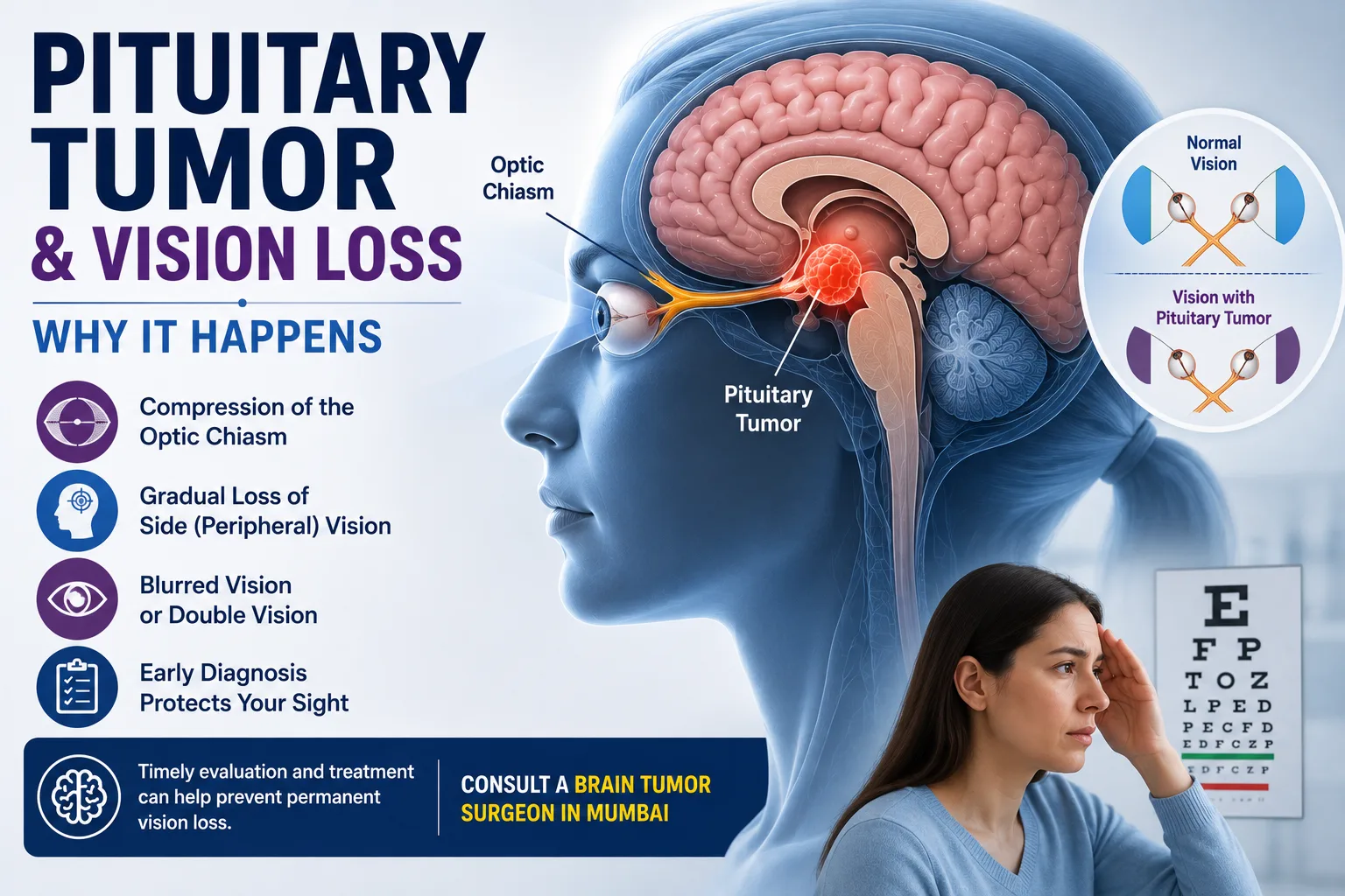
Vision may change gradually enough that a person does not immediately realise something is wrong. They may start bumping into objects at the side, struggle to notice vehicles approaching from either direction, or feel that their peripheral vision is narrowing. Pituitary tumor vision loss can develop when an enlarging tumor grows upward from the pituitary gland and presses on the optic nerves or optic chiasm—the structure where visual nerve fibres from both eyes cross.
Not every pituitary tumor affects eyesight. Small tumors may cause hormonal symptoms without any visual problems, while larger tumors are more likely to affect nearby structures. Recognised pressure-related symptoms include loss of side vision, reduced eyesight and double vision.
Understanding the pattern of vision change, together with formal eye testing and a pituitary MRI, helps doctors determine whether the tumor is affecting the visual pathways.
Why Can a Pituitary Tumor Cause Vision Loss?
Pituitary tumor vision loss usually occurs because the pituitary gland sits directly below important visual pathways. If a tumor grows upward, it can compress the optic chiasm or optic nerves. This pressure can interfere with nerve signals travelling from the eyes to the brain, causing peripheral visual-field loss, blurred vision or reduced visual clarity.
The pituitary gland sits in a small bony space at the base of the brain called the sella turcica. Immediately above it lies the optic chiasm.
The optic nerves carry information from each eye toward the brain. Some of these nerve fibres cross at the optic chiasm before continuing toward areas of the brain responsible for processing vision.
A larger pituitary adenoma can grow beyond the sella and upward toward this structure. This is why the direction of tumor growth can matter as much as its overall size.
What Is Optic Chiasm Compression?
Optic chiasm compression means that a mass is pressing on the area where fibres from the two optic nerves partially cross.
Pituitary adenomas are an important cause of optic chiasm compression in adults. The resulting visual-field problem commonly affects the outer, or temporal, field of vision in both eyes.
This pattern is called bitemporal hemianopia.
In simple terms, a person may continue seeing reasonably well straight ahead but gradually lose awareness of things approaching from either side.
That can create practical problems such as:
- Missing objects located toward either side
- Bumping into doorframes or furniture
- Difficulty noticing cars approaching from the side
- Feeling less confident while walking in busy environments
- Difficulty with activities that depend on a wide visual field
Visual-field loss may initially be asymmetrical or incomplete, so patients do not always recognise it themselves.
What Pituitary Tumor Eye Symptoms Should You Notice?
Pituitary tumor eye symptoms depend on the tumor’s size, direction of growth and which visual structures are compressed.
Possible symptoms include:
- Loss of peripheral or side vision
- Blurred vision
- Reduced visual sharpness
- Difficulty seeing objects toward either side
- Double vision
- Unequal vision between the two eyes
- Drooping of an eyelid in some cases
- Difficulty moving an eye normally in less common situations
Mayo Clinic lists loss of side vision and double vision among pressure-related symptoms of larger pituitary tumors.
Visual problems may develop slowly. Someone may therefore adapt without realising how much peripheral vision has been lost.
Family members may occasionally notice practical changes first—for example, the person repeatedly failing to notice things positioned at the edge of their visual field.
Can a Pituitary Tumor Cause Blurred Vision?
Yes. Pituitary tumor blurred vision can occur, although blurred vision has many possible causes unrelated to the pituitary gland.
A larger pituitary tumor may affect the optic nerves or chiasm enough to reduce visual clarity. Some patients primarily notice loss of peripheral vision, while others describe general blurring or difficulty seeing clearly.
Blurred vision alone is not enough to diagnose a pituitary adenoma.
More common causes include refractive errors, cataract, dry eyes, retinal disease and several other eye conditions. The pattern becomes more concerning for a neurological cause when blurred vision is progressive, accompanied by visual-field loss, occurs with double vision, or is associated with headaches or known pituitary abnormalities.
Can a Pituitary Adenoma Cause Double Vision?
Yes, although double vision is less typical than loss of peripheral vision.
Pituitary adenoma vision problems can occasionally include double vision if the tumor extends laterally toward an area called the cavernous sinus. Several nerves responsible for controlling eye movement travel through or close to this region.
Compression of these nerves can interfere with the coordinated movement of the eyes, producing double vision.
A patient may notice:
- Two images of one object
- Difficulty keeping images aligned
- Double vision in a particular direction
- An eyelid beginning to droop
- Abnormal eye movement
New double vision deserves medical assessment, particularly when it develops suddenly or occurs with a severe headache.
Does Every Large Pituitary Tumor Cause Vision Loss?
No.
A larger tumor is more likely to reach nearby structures, but size alone does not determine visual symptoms.
Doctors also consider:
- Direction of tumor growth
- Distance between the tumor and optic chiasm
- Degree of optic nerve compression
- How quickly the lesion has enlarged
- Individual anatomical differences
- Duration of compression
A tumor may grow in a direction that produces hormonal or other pressure symptoms before vision becomes affected.
Conversely, a tumor situated directly beneath the optic chiasm may threaten visual function even when other symptoms are relatively limited.
This is why treatment decisions are based on the actual MRI appearance and visual testing rather than tumor size alone.
How Do Doctors Check Whether Vision Loss Is From a Pituitary Tumor?
Evaluation usually involves more than asking whether the patient can read an eye chart.
Formal Visual-Field Testing
A visual-field test measures how much of the surrounding area a person can see while looking straight ahead.
This is particularly useful because pituitary tumors can affect side vision before patients notice a major reduction in central vision. Eye examinations are routinely used to assess the side vision of people with pituitary tumors.
Testing may show a characteristic bitemporal pattern when the optic chiasm is compressed.
Visual-field measurements can also provide a baseline for comparison after treatment.
Visual Acuity and Eye Examination
Doctors may assess how clearly each eye sees and examine the eyes for other causes of visual loss.
An ophthalmologist or neuro-ophthalmologist may become involved when the visual findings need more detailed evaluation.
Pituitary MRI
A dedicated pituitary MRI provides detailed images of the gland, tumor and surrounding structures.
Doctors examine:
- Tumor dimensions
- Whether it extends above the pituitary gland
- Contact with or compression of the optic chiasm
- Extension toward the cavernous sinus
- Relationship with nearby blood vessels
- Appearance after contrast administration
Larger adenomas can enlarge the gland and press on nearby structures involved in vision.
Hormone Testing
Pituitary tumors can affect hormone production as well as vision.
Blood tests may therefore include prolactin, cortisol-related testing, thyroid hormones, growth hormone-related tests and reproductive hormones depending on the clinical situation.
The hormone profile matters because the exact tumor type can change the treatment plan.
Does Vision Loss Mean a Pituitary Tumor Needs Surgery?
Not always, but confirmed visual compression is an important reason for specialist treatment assessment.
Surgery is commonly considered when a pituitary tumor presses on the optic nerves or is already affecting vision. Other reasons can include hormone overproduction, continued tumor growth or compression of normal pituitary tissue.
The most common surgical route is transsphenoidal surgery. This allows the surgeon to reach the pituitary region through the nose and sphenoid sinus rather than through a large opening in the skull.
The aim may include:
- Relieving pressure on the optic pathways
- Removing as much tumor as safely possible
- Controlling tumor growth
- Obtaining tissue when diagnosis requires it
- Managing hormone-producing tumors when surgery is appropriate
Surgery is not the first treatment for every pituitary tumor.
Prolactinomas Are an Important Exception
A prolactinoma is a pituitary tumor that produces excessive prolactin.
These tumors are commonly treated first with medicines known as dopamine agonists. The medicines can reduce prolactin levels and often shrink the tumor, which can also relieve pressure-related vision problems.
Surgery may be considered if medication is ineffective, unsuitable or poorly tolerated, or when other clinical circumstances require it.
Other pituitary tumor types have different treatment pathways.
Will Vision Return After Pituitary Tumor Treatment?
Vision can improve after the pressure on the visual pathways is relieved, but the degree of recovery cannot be guaranteed.
Several factors may influence recovery, including:
- Severity of vision loss before treatment
- Duration of optic nerve or chiasm compression
- Degree of visual-field damage
- Tumor size and location
- Health of the optic nerves
- Treatment used
Long-standing compression can damage nerve fibres. This is one reason progressive vision loss should be assessed rather than simply watched without medical guidance.
Even after treatment, visual-field testing and follow-up MRI may be needed to document recovery and monitor the remaining pituitary region.
When Is Pituitary Tumor Vision Loss an Emergency?
Most pituitary adenomas grow gradually, so their visual symptoms usually develop over time. One situation is very different: pituitary apoplexy.
Pituitary apoplexy occurs when sudden bleeding or loss of blood supply develops within a pituitary tumor. It can cause rapid enlargement and acute pressure on nearby structures.
Seek urgent medical care for symptoms such as:
- Sudden, extremely severe headache
- Sudden vision loss
- Rapid worsening of vision
- New double vision
- Vomiting
- New confusion
- Unusual drowsiness or loss of consciousness
Pituitary apoplexy requires immediate medical care.
Sudden visual loss can also result from several other eye and neurological emergencies. It should never be assumed to be due to a known pituitary tumor without prompt assessment.
Neurosurgical review is particularly relevant when MRI shows a pituitary tumor touching or compressing the optic chiasm, when formal visual testing demonstrates progressive loss, or when surgery is being considered.
A specialist assessment may review:
- The actual MRI images
- Degree of optic chiasm compression
- Visual-field test results
- Visual acuity
- Tumor size and direction of growth
- Hormonal test results
- Whether medication should be tried first
- Whether surgery is indicated
- Potential surgical risks and treatment goals
Patients with a pituitary lesion affecting vision may seek evaluation from a brain tumor surgeon in Mumbai to understand whether neurosurgical treatment has a role.
Dr. Mazda K. Turel can review the scan alongside visual and hormonal findings before discussing treatment. Pituitary tumors are often best assessed through a multidisciplinary approach involving neurosurgery, endocrinology and ophthalmology or neuro-ophthalmology.
Conclusion
Pituitary tumor vision loss usually develops because an enlarging tumor reaches the visual pathways above the pituitary gland, particularly the optic chiasm. Loss of side vision is a characteristic pattern, but patients may also experience blurred vision, reduced visual clarity or double vision.
These symptoms should not be used for self-diagnosis. Many eye and neurological disorders can produce similar changes.
A dedicated pituitary MRI, formal visual-field examination and hormone testing can show whether a pituitary tumor is responsible and help determine treatment. If the optic nerves are being compressed, timely specialist assessment matters because prolonged pressure can affect the potential for visual recovery.
Sudden severe headache combined with abrupt vision loss, double vision, vomiting or confusion requires urgent medical assessment because pituitary apoplexy is a medical emergency.
Frequently Asked Questions
What type of vision loss does a pituitary tumor cause?
A classic pattern is loss of the outer visual field in both eyes, known as bitemporal hemianopia. It occurs when a pituitary tumor compresses the optic chiasm. The pattern is not identical in every patient, however. Some people develop asymmetric field loss, reduced visual acuity, blurred vision or other abnormalities depending on the tumor’s direction and extent.
Can pituitary tumor vision loss affect only one eye?
It can. Although optic chiasm compression commonly affects the visual fields of both eyes, an asymmetrical tumor can affect one optic nerve or one side of the chiasm more than the other. This may create unequal visual symptoms. Formal visual-field testing and MRI help determine which part of the visual pathway is being compressed.
Is pituitary tumor blurred vision permanent?
Not necessarily. Pituitary tumor blurred vision may improve when pressure on the visual pathways is relieved, but recovery varies between patients. Long-standing or severe optic nerve damage can limit improvement. The duration of symptoms, severity of compression and health of the optic nerves all influence the potential for visual recovery.
Can a small pituitary tumor affect eyesight?
Small pituitary tumors are less likely to cause compression-related visual symptoms because they usually remain within the pituitary region. However, treatment decisions should not be made from size alone. Doctors look at the tumor’s exact position and direction of growth, hormone activity, symptoms and its relationship with the optic nerves and chiasm.
Does loss of peripheral vision always mean a pituitary tumor?
No. Peripheral visual-field loss has several possible eye and neurological causes. Pituitary tumors are one recognised cause because they can compress the optic chiasm. If unexplained peripheral vision loss is detected, an eye specialist or neurologist may recommend further investigation based on the visual-field pattern and examination findings.
Can prolactinoma cause vision loss?
Yes, particularly if a prolactinoma becomes large enough to press on the optic nerves or optic chiasm. Larger prolactinomas can produce headaches and vision problems. Unlike many other pituitary tumors, prolactinomas are usually treated first with medicines that lower prolactin and can shrink the tumor.
Is surgery always required when the optic chiasm is compressed?
Not in every situation. Treatment depends partly on the tumor type and severity of visual compromise. A prolactinoma may shrink substantially with medication, for example. Other tumors producing significant visual-field loss may be treated surgically. The decision requires review of MRI findings, visual testing, hormone results and the patient’s overall clinical circumstances.